
Maternal and Fetal Mitochondrial Gene Dysregulation in Hypertensive Disorders of Pregnancy
- Details
- Published on 04 August 2023


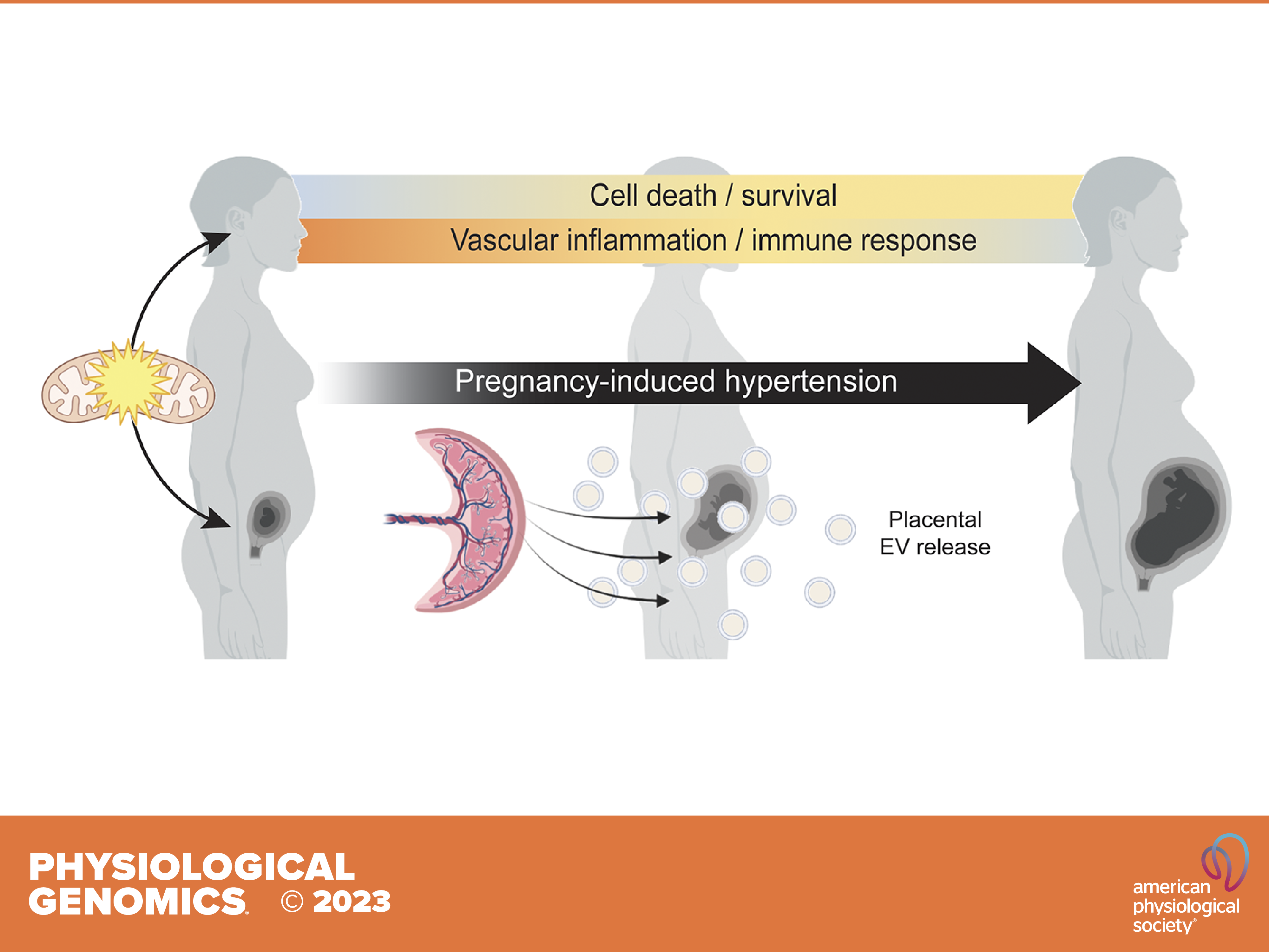
Mitochondrial dysfunction has been implicated in pregnancy-induced hypertension (PIH). The role of mitochondrial gene dysregulation in PIH, and consequences for maternal-fetal interactions, remain elusive.
In their new study, Nicole R. Phillips (University of North Texas Health Science Center), Styliani Goulopoulou (Loma Linda University) and their colleagues investigated mitochondrial gene expression and dysregulation in maternal and placental tissues from pregnancies with and without PIH.
Further, they measured circulating mitochondrial DNA (mtDNA) mutational load, an index of mtDNA integrity. Differential gene expression analysis followed by Time Course Gene Set Analysis (TcGSA) was conducted on publicly available high throughput sequencing transcriptomic data sets. Mutational load analysis was carried out on peripheral mononuclear blood cells from healthy pregnant individuals and individuals with preeclampsia.
Thirty mitochondrial differentially expressed genes (mtDEGs) were detected in the maternal cell-free circulating transcriptome, whereas nine were detected in placental transcriptome from pregnancies with PIH. In PIH pregnancies, maternal mitochondrial dysregulation was associated with pathways involved in inflammation, cell death/survival, and placental development, whereas fetal mitochondrial dysregulation was associated with increased production of extracellular vesicles (EVs) at term. Mothers with preeclampsia did not exhibit a significantly different degree of mtDNA mutational load.
The reported findings support the involvement of maternal mitochondrial dysregulation in the pathophysiology of PIH and suggest that mitochondria may mediate maternal-fetal interactions during healthy pregnancy.
In summary, this study identifies aberrant maternal and fetal expression of mitochondrial genes in pregnancies with gestational hypertension and preeclampsia. Mitochondrial gene dysregulation may be a common etiological factor contributing to the development of de novo hypertension in pregnancy-associated hypertensive disorders.
Image credits: Ricci et al. Physiological Genomics (2023)
Targeting Mitochondria 2023, this October, will shed light on the latest discoveries related to mitochondria. Submit a related abstract.
Media contact:
World Mitochondria Society
This email address is being protected from spambots. You need JavaScript enabled to view it.
+33-1-5504-7755
Targeting Mitochondria 2023 Congress
October 11-13, 2023 - Berlin, Germany
wms-site.com
Freiburg research team casts light on signal-dependent formation of mitochondria
- Details
- Published on 03 August 2023
Scientists reveal the transport of positively charged signal sequences through negatively charged groove
Known as the power plant of the cell, mitochondria are essential to human metabolism. Human mitochondria consist of 1,300 different proteins and two fatty biomembranes. The vast majority of mitochondrial proteins are produced with a cleavable transport signal and have to be actively transported into the mitochondria. Using biochemical and cell-biology experiments, a team of researchers have now shown for the first time precisely how mitochondrial proteins with signal sequences are imported into the mitochondria via a negatively charged, unique groove. Headed by Prof. Dr. Nils Wiedemann and Prof. Dr. Carola Hunte from the Medical Faculty at the University of Freiburg, and Prof. Dr. Martin van der Laan from the University of Saarland the work was carried out at the University of Freiburg’s Cluster of Excellence CIBSS – Centre for Integrative Biological Signalling Studies. Their results have been published in the science journal Nature.
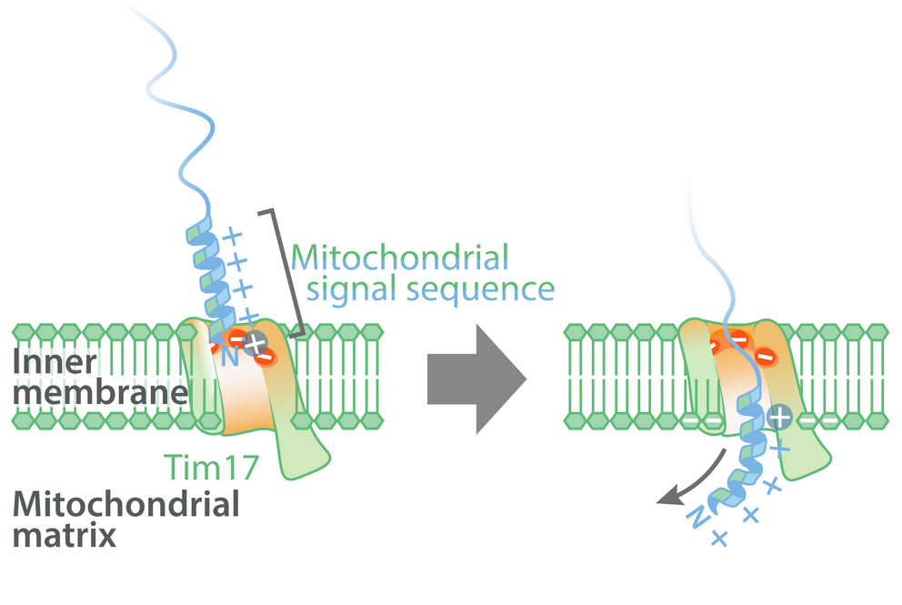
Model of the import of mitochondrial proteins with a signal sequence across the mitochondrial inner membrane at the Tim17 groove. Illustration: Laura Fielden
Transport mechanism is an important building block
“Forty years after the discovery of mitochondrial signal sequences, our experiments have now revealed the precise mechanism by which proteins are transported and the power stations of our cells are gradually built up,” says Wiedemann. “Information about the transport mechanism for mitochondrial proteins is an important component in basic cellular research.” Malfunctions of over 500 mitochondrial proteins cause a variety of diseases, so research into the mitochondria is hugely important to medicine.
It was already known that mitochondrial proteins were imported using the signal sequence translocase of the inner membrane (TIM) into the mitochondrial matrix. The two vital core subunits of this translocase are Tim17 and Tim23. Until now, it was assumed that mitochondrial proteins with signal sequences were transported across the inner membrane via a water-filled Tim23 channel. However, recent artificial intelligence-based structural predictions indicate that Tim23 does not form a channel. The research team has now been able to prove that mitochondrial proteins with signal sequences are actually imported into the mitochondria via a groove in the Tim17 protein.
Negatively charged groove in Tim17-protein
Most proteins that are transported into the mitochondria contain a complex molecular signal sequence which is positively charged and water-soluble on one side, and on the opposite side has fat-soluble molecular residues. In contrast to the positively charged side of the transport signal, the groove of Tim17 contains a strongly negatively charged region which is present in all Tim17 proteins, from yeast to humans.
The lead authors of the study, Dr. Laura Fielden and Dr. Jakob Busch from the Institute of Biochemistry and Molecular Biology at the University of Freiburg, employed functional in vitro transport experiments using chemically-marked proteins with isolated mitochondria to show that the negative charges in the groove of Tim17 interact with the positively charged signal sequences and are therefore essential to the transportation of mitochondrial proteins. Meanwhile the fat-soluble side of the mitochondrial signal sequences is aligned with the lipid membrane, enabling the transport of signal sequences at the interface between Tim17 and the mitochondrial inner membrane.
Basis for further research
“Now we have clarified this fundamental mechanism of mitochondrial proteins with a signal sequence at the interface to the biomembrane, we can understand why mitochondrial signal sequences have one positively charged side and one fat-soluble side and need this for their transportation,” Fielden explains, emphasising the importance of the results which can now serve as a basis for further research into mitochondria.
Source: Universität Freiburg
Targeting Mitochondria 2023, this October, will shed light on the latest discoveries related to mitochondria. Submit a related abstract.
Media contact:
World Mitochondria Society
This email address is being protected from spambots. You need JavaScript enabled to view it.
+33-1-5504-7755
Targeting Mitochondria 2023 Congress
October 11-13, 2023 - Berlin, Germany
wms-site.com
Study Shows Mitochondrial Transplantation Effective in Reversing Damage to Kidneys and Kidney Cells
- Details
- Published on 28 July 2023
According to the National Kidney Foundation, more than 100,000 Americans are waiting for a kidney transplant, and the demand for donated kidneys far exceeds the supply. In fact, only 25,498 kidney transplants were performed in 2022, and kidney disease impacts 37 million people in the U.S.
But a new preclinical study, led by scientists at Wake Forest University School of Medicine, shows that a new technology called mitochondrial transplantation holds promise as a potential therapy that could change the kidney transplant landscape.
The study findings appear online in Annals of Surgery.
Mitochondrial transplantation is a regenerative medicine technology where healthy mitochondria are taken from cultured cells or tissue from organ donors and then injected into a diseased or damaged tissue or organ. Mitochondria produce the energy needed for a cell to function.
“Our study shows that this technology could transform renal transplant medicine,” said Giuseppe Orlando, M.D., Ph.D., an associate professor of surgery at Wake Forest University School of Medicine, a transplant surgeon at Atrium Health Wake Forest Baptist and principal investigator of the study. “Here, we provide evidence that mitochondrial transfer lessens the damage that renal cells or the kidneys may suffer from disease or injury.”
For the study, the research team collaborated with the University of Turin in Italy where scientists conducted preliminary tests in cultures of human proximal tubular cells, which are found in the kidneys and play an important in removing toxins. When the damaged cells were exposed to healthy mitochondria, cellular energy increased, and toxicity decreased.
“Essentially, the mitochondrial transplant was successful in reducing stress in the damaged kidney cells,” said Orlando, who is also a researcher at Wake Forest Institute for Regenerative Medicine.
Additional research by the Wake Forest team found that kidneys injected with healthy mitochondria showed signs of recovery.
Orlando said these results are significant because in the U.S., 20% of the kidneys procured for transplantation are eventually discarded because they are too damaged, and this potential new treatment may help.
“Based on these preliminary findings, I’m optimistic that mitochondrial transplantation could one day increase the number of transplantable organs,” Orlando said.
Orlando said this is especially true in a new type of organ donation called “uncontrolled donation after cardiac death,” an area of active research. In this setting, the kidneys do not receive adequate blood supply, and mitochondria and the kidneys are damaged. Orlando said mitochondrial transplantation could enable surgeons to repair and eventually transplant these organs, potentially saving thousands of lives. It has been estimated that this type of donation may make 20,000 new kidneys suitable for transplants each year in the U.S., Orlando said.
The next step is to validate these findings in a small pilot study.
This study was supported by the National Center for Advancing Translational Sciences, National Institutes of Health Grant No. UL1TR001420 and the Italian Ministry of Health and Research.
Source: Atrium Health Wake Forest Baptist
Image Credits: Image Credits: WangXiNa on Freepik
Dr. Orlando will be joining Targeting Mitochondria 2023 to give a talk entitled "Repairing Marginal Kidneys With Mitochondrial Transplantation: A New Powerful Tissue Engineering Tool That Will Change the Transplant Landscape". Submit a related abstract.
Media contact:
World Mitochondria Society
This email address is being protected from spambots. You need JavaScript enabled to view it.
+33-1-5504-7755
Targeting Mitochondria 2023 Congress
October 11-13, 2023 - Berlin, Germany
wms-site.com
Hydrogen Sulfide: A Promising Healthy ageing Therapeutic When Specifically Targeted Within Cells
- Details
- Published on 01 August 2023

News Release, World Mitochondria Society, Berlin - Germany – August 1, 2023
Future therapies to help people live healthy lives for longer could be developed from drugs that release tiny amounts of the gas hydrogen sulfide (H2S), new research has indicated.
A study from the University of Exeter, funded by the US Army and charity The United Mitochondrial Disease Foundation, found that targeting tiny amounts of H2S to specific areas of cells in adult worms using a H2S-releasing molecule called AP39, greatly improved health and activity as they aged. The research, published in PNAS, concludes that targeting H2S specifically to the energy-generating machinery of cells (mitochondria) could one day be used as a healthy aging therapeutic.
The research team administered AP39 to some worms from birth, and to others after reaching adulthood. They found that this compound improved the integrity of mitochondria – the “power house” of cells, which produces our cells’ energy, and kept the worms’ muscles active and moving, even well into old age, and when given mid-way through their life-course.
A number of age-related conditions are linked to loss of mitochondrial function, including natural ageing, neurodegenerative diseases such as Parkinson’s and Alzheimer’s as well as muscular dystrophy and primary mitochondrial diseases.
The team also found a group of proteins that regulated how genes are expressed in ageing (transcription factors). There transcription factors were found to be specifically targeted by H2S. This insight may identify new targets for therapy in ageing and age-related conditions, particularly conditions affecting muscle.
Senior author Professor Tim Etheridge, of the University of Exeter, said: “Worms are a powerful genetic tool to study human health and disease and offer a strong platform to quickly identify new potential therapeutics. Diseases related to ageing take a huge toll on society. Our results indicate that H2S, administered to specific parts of the cell in tiny quantities, could one day be used to help people live healthier for longer
In previous research, the team had found that they could successfully target skeletal muscle with H2S in worms, and the new paper represents the first time this technique has been applied to natural ageing.
The University of Exeter has assigned the underlying technology to its spin-out MitoRx Therapeutics, which has developed next generation compounds with much better drug characteristics as potential medicines to combat diseases of ageing including neurodegenerative disorders such as Huntington’s disease as well as rare childhood conditions such as muscular dystrophy.
Co-author Professor Matt Whiteman, from the University of Exeter, said: “This study is not about extending life – it’s about living healthier lives well into older age. This could have huge benefits to society. We’re excited to see this research move to the next stages over the coming years, and hope it will one day form the basis of new treatments which we have the potential to develop with MitoRx.”
“We saw a small extension of lifespan in the worms that were targeted with H2S, and what’s unique here is that we extended healthspan – or the time they lived healthy lives. The worms still died, albeit later than normally expected, but they died very active and with young physiology.”
The paper is entitled ‘Mitochondrial sulfide promotes lifespan and healthspan through distinct mechanisms in developing versus adult treated Caenorhabditis elegans, and is published in PNAS.
Source: University of Exeter.
Targeting Mitochondria 2023, this October, will dedicate a whole session to "Mitochondria & Longevity: Towards Expanding Life Span". Submit a related abstract.
Media contact:
World Mitochondria Society
This email address is being protected from spambots. You need JavaScript enabled to view it.
+33-1-5504-7755
Targeting Mitochondria 2023 Congress
October 11-13, 2023 - Berlin, Germany
wms-site.com
Inflammation Discovery Could Slow Aging, Prevent Age-Related Diseases: Strategic Role of Mitochondria
- Details
- Published on 25 July 2023

A new discovery from School of Medicine researchers could let us slow the clock on aging and may help us prevent many diseases that plague us in our later years.
School of Medicine researchers have discovered a key driver of chronic inflammation that accelerates aging. That finding could let us slow the clock to live longer, healthier lives, and may allow us to prevent age-related conditions such as deadly heart disease and devastating brain disorders that rob us of our faculties.
So what drives this harmful inflammation? The answer is improper calcium signaling in the mitochondria of certain immune cells. Mitochondria are the power generators in all cells, and they rely heavily on calcium signaling.
The UVA Health researchers, led by Bimal N. Desai, PhD, found that the mitochondria in the immune cells called macrophages lose their ability to take up and use calcium with age. This, the researchers show, leads to chronic inflammation responsible for many of the ailments that afflict our later years.
The researchers believe that increasing calcium uptake by the mitochondrial macrophages could prevent the harmful inflammation and its terrible effects. Because macrophages reside in all organs of our bodies, including the brain, targeting such “tissue-resident macrophages” with appropriate drugs may allow us to slow age-associated neurodegenerative diseases.
“I think we have made a key conceptual breakthrough in understanding the molecular underpinnings of age-associated inflammation,” said Desai, of UVA’s Department of Pharmacology and UVA’s Carter Immunology Center. “This discovery illuminates new therapeutic strategies to interdict the inflammatory cascades that lie at the heart of many cardiometabolic and neurodegenerative diseases.”
The Inflammation of Aging – ‘Inflammaging’
Macrophages are white blood cells that play critical roles in our immune systems and, in turn, our good health. They swallow up dead or dying cells, allowing our bodies to remove cellular debris, and patrol for pathogens and other foreign invaders. In this latter role, they act as important sentries for our immune systems, calling for help from other immune cells as needed.
Scientists have known that macrophages become less effective with age, but it has been unclear why. Desai’s new discovery suggests answers.
Desai and his team say their research has identified a “keystone” mechanism responsible for age-related changes in the macrophages. These changes, the scientists believe, make the macrophages prone to chronic, low-grade inflammation at the best of times. And when the immune cells are confronted by an invader or tissue damage, they can become hyperactive. This drives what is known as “inflammaging” – chronic inflammation that drives aging.
Further, the UVA Health scientists suspect that the mechanism they have discovered will hold true not just for macrophages but for many other related immune cells generated in the bone marrow. That means we may be able to stimulate the proper functioning of those cells as well, potentially giving our immune systems a big boost in old age, when we become more susceptible to disease.
Next Steps
Fixing “inflammaging” won’t be as simple as taking a calcium supplement. The problem is not a shortage of calcium so much as the macrophages’ inability to use it properly. But Desai’s new discovery has pinpointed the precise molecular machinery involved in this process, so we should be able to discover ways to stimulate this machinery in aging cells.
“This highly interdisciplinary research effort, at the interface of computational biology, immunology, cell biology and biophysics, wouldn’t have been possible without the determination of Phil Seegren, the graduate student who spearheaded this ambitious project,” Desai said. “Now, moving forward, we need an equally ambitious effort to figure out the wiring that controls this mitochondrial process in different types of macrophages and then manipulate that wiring in creative ways for biomedical impact.”
Aging Findings Published
The researchers have published their findings in the scientific journal Nature Aging. The article is open access, meaning it is free to read.
The research team consisted of Philip V. Seegren, Logan R. Harper, Taylor K. Downs, Xiao-Yu Zhao, Shivapriya B. Viswanathan, Marta E. Stremska, Rachel J. Olson, Joel Kennedy, Sarah E. Ewald, Pankaj Kumar and Desai. The scientists reported that they have no financial interests in the work.
The research was supported by the National Institutes of Health, grants AI155808, GM108989, GM138381, P30 CA044579 and T32 GM007055-46, and by the Owens Family Foundation.
Source: UVA Health
Targeting Mitochondria 2023 will introduce you to the latest research on mitochondria and longevity. Submit a related abstract.
Media contact:
World Mitochondria Society
This email address is being protected from spambots. You need JavaScript enabled to view it.
+33-1-5504-7755
Targeting Mitochondria 2023 Congress
October 11-13, 2023 - Berlin, Germany
wms-site.com


