
Mitochondrial Dysfunction in Cell Senescence and Aging
- Details
- Published on 27 January 2023


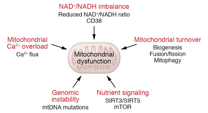
Mechanisms that can cause mitochondrial dysfunction.
News Release, World Mitochondria Society, Berlin - Germany – January 30, 2023
Mitochondrial dysfunction and cell senescence are hallmarks of aging and are closely interconnected. Mitochondrial dysfunction, operationally defined as a decreased respiratory capacity per mitochondrion together with a decreased mitochondrial membrane potential, typically accompanied by increased production of oxygen free radicals, is a cause and a consequence of cellular senescence and figures prominently in multiple feedback loops that induce and maintain the senescent phenotype.
Here, Miwa et al. summarized pathways that cause mitochondrial dysfunction in senescence and aging and discussed the major consequences of mitochondrial dysfunction and how these consequences contribute to senescence and aging. They also highlighted the potential of senescence-associated mitochondrial dysfunction as an antiaging and antisenescence intervention target, proposing the combination of multiple interventions converging onto mitochondrial dysfunction as novel, potent senolytics.
© Image - Miwa et al., J Clin Invest. 2022 Jul.
Targeting Mitochondria 2023 will dedicate a whole session to mitochondria & longevity. Access Sessions 2023.
Media contact:
World Mitochondria Society
This email address is being protected from spambots. You need JavaScript enabled to view it.
+33-1-5504-7755
Targeting Mitochondria 2023 Congress
October 11-13, 2023 - Berlin, Germany
wms-site.com
Mitochondria Transplant Therapy for Injured Skeletal Muscle
- Details
- Published on 12 January 2023
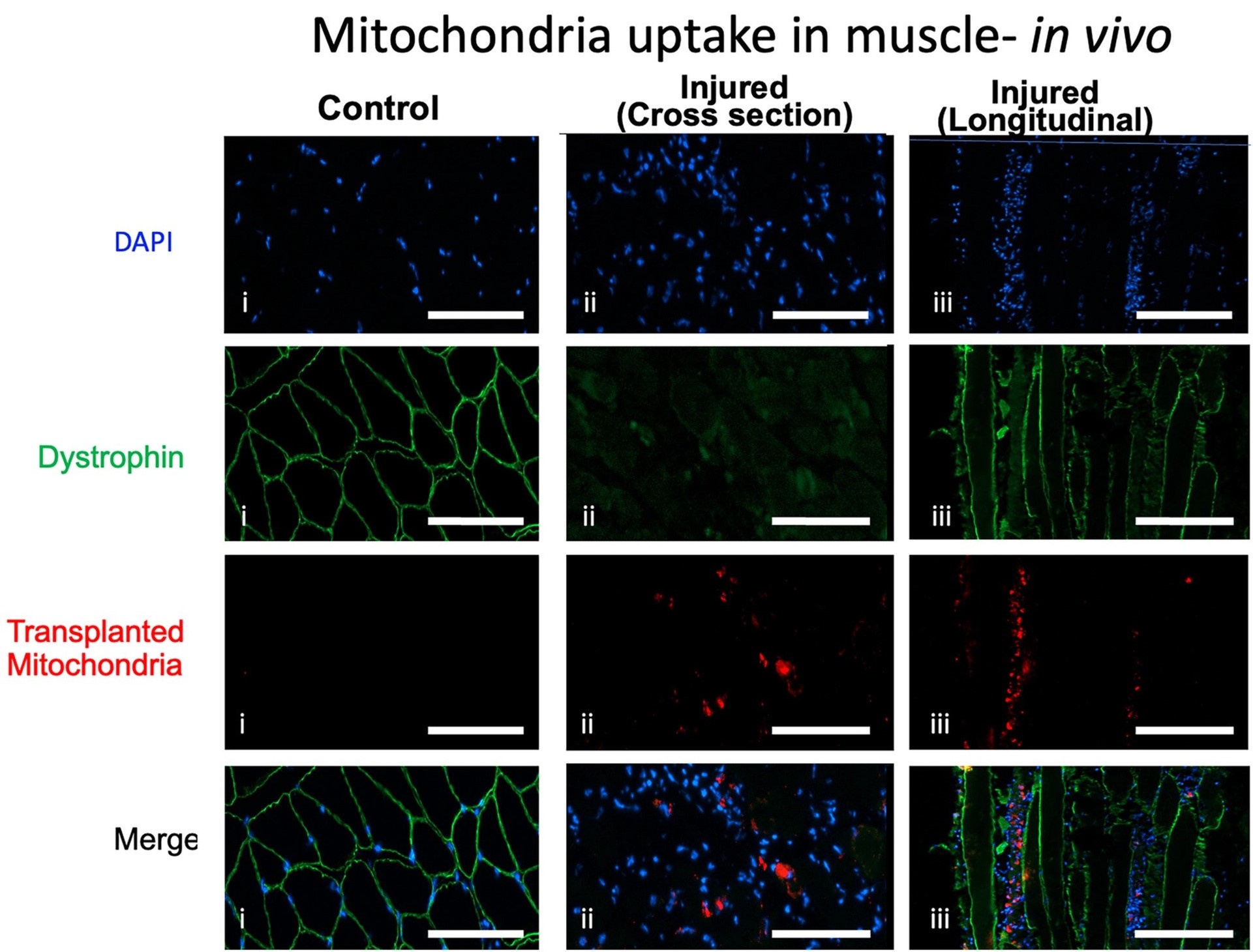
Uptake of donor mitochondria in myoblasts, myotubes and myofibres.
News Release, World Mitochondria Society, Berlin - Germany – January 12, 2023
- BaCl2 was injected into the gastrocnemius muscle of one limb of 8–12-week-old C57BL/6 mice to induce damage without injury to the resident stem cells.
- The contralateral gastrocnemius muscle was injected with phosphate-buffered saline (PBS) and served as the non-injured intra-animal control.
- Mitochondria were isolated from donor mice.
- Donor mitochondria were suspended in PBS or PBS without mitochondria (sham treatment) and injected into the tail vein of BaCl2 injured mice 24 h after the initial injury.
- Muscle repair was examined 7, 14 and 21 days after injury.
MTT did not increase systemic inflammation in mice. Muscle mass 7 days following injury was 21.9 ± 2.1% and 17.4 ± 1.9% lower (P < 0.05) in injured as compared with non-injured intra-animal control muscles in phosphate-buffered saline (PBS)- and MTT-treated animals, respectively.
Maximal plantar flexor muscle force was significantly lower in injured as compared with uninjured muscles of PBS-treated and MTT-treated mice, but the reduction in force was not different between the experimental groups. The percentage of collagen and other non-contractile tissue in histological muscle cross sections, was significantly greater in injured muscles of PBS-treated mice compared with MTT-treated mice 7 days after injury.
Muscle wet weight and maximal muscle force from injured MTT-treated mice had recovered to control levels by 14 days after the injury. However, muscle mass and force had not improved in PBS-treated animals by 14 days after injury.
By 21 days following injury, PBS-treated mice had fully restored gastrocnemius muscle mass of the injured muscle to that of the uninjured muscle, although maximal plantar flexion force was still lower in injured/repaired gastrocnemius as compared with uninjured intra-animal control muscles.
© Image - Alway et al., Journal of Cachexia, Sarcopenia and Muscle (2023)
Media contact:
World Mitochondria Society
This email address is being protected from spambots. You need JavaScript enabled to view it.
+33-1-5504-7755
Targeting Mitochondria 2023 Congress
October 11-13, 2023 - Berlin, Germany
wms-site.com
Mitochondria Targeted Near-Infrared Aggregation-Induced Emission for Photodynamic Ablation of Liver Cancer Cells
- Details
- Published on 06 September 2022
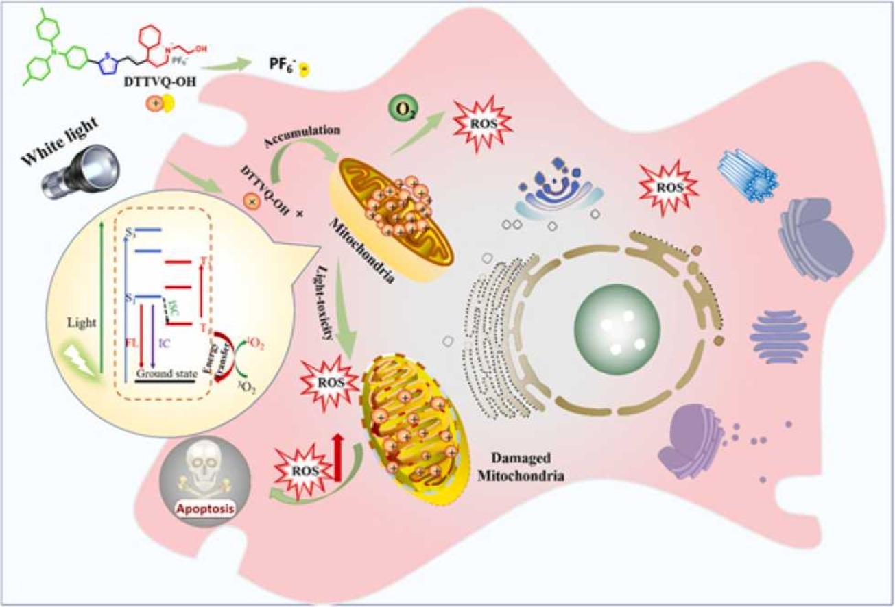
Multifunctional nano-photosensitizer was used for mitochondrial-targeting photodynamic ablation of liver cancer cells.
News Release, World Mitochondria Society, Berlin - Germany – September 6, 2022
The aggregation-induced emission photosensitizers (AIE-PSs) manifest a multitude of notable superiorities in terms of high specificity to organelles, high-efficient singlet oxygen (1O2) generation as well as enhanced fluorescence intensity, which provides a feasible approach to overcome the problems such as the insufficient generation of reactive oxygen species (ROS) caused by grave aggregation-induced quenching (ACQ) and the lack of specific targeting existing in traditional PSs, but extremely challenging.
Herein, a series of near-infrared (NIR) AIE luminogens (AIEgens) for targeting mitochondrial was devised and synthesized by regulating the D-A intensity assembly molecular engineering, which fabricating a progressively stronger intermolecular charge transfer (ICT) state to accelerate highly effective intersystem crossing (ISC) of excited electrons by the synergistic effect of thiophene and quinolinium.
- The optimal NIR AIE-PS (DTTVQ-OH) revealed excellent photostability, biocompatibility, precise mitochondria targeting, extremely high generation yield of 1O2 and superior phototoxicity in living HepG2 cells.
- Apoptosis assay and cell migration experiment further demonstrated that DTTVQ-OH could efficaciously restrain cell proliferation and induce/speed up cancer cell death.
- DTTVQ-OH could selectively distinguish cancer cells and normal cells by difference of fluorescence intensity in high resolution without the assist of any extra targeting ligands.
As a consequence, this work provides a rational and practicable strategy for the specific targeted molecular engineering of AIE-PSs, which gives impetus to the development of fluorescence imaging-guided photodynamic therapy fields.
© Image -Xue et al., Sensors and Actuators B: Chemical 2022
Targeting Mitochondria 2022 will dedicate a whole session to "Translational Therapies - Focus on Infrared Therapies". Submit a related abstract.
Media contact:
World Mitochondria Society
This email address is being protected from spambots. You need JavaScript enabled to view it.
+33-1-5504-7755
Targeting Mitochondria 2022 Congress
October 26-28, 2022 - Berlin, Germany
wms-site.com
Researchers Tailor Blood Pressure Drug Delivery Directly to Cells’ ‘Power Plants’
- Details
- Published on 22 September 2022
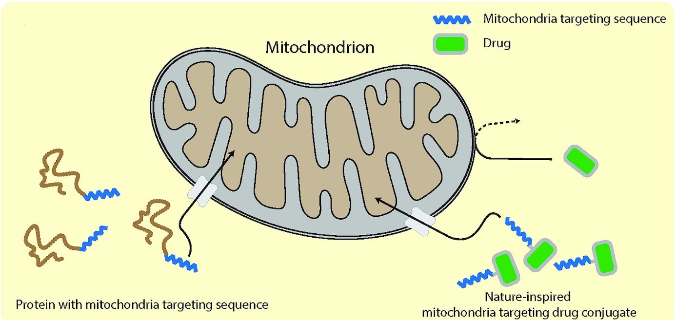
Schematic illustration of the design, synthesis, and mechanism of uptake of the mitochondrially targeted Losartan.
News Release, World Mitochondria Society, Berlin - Germany – September 6, 2022
In a study using lab-grown cells, Johns Hopkins Medicine researchers specializing in aging report they have successfully delivered a common blood pressure drug directly to the inner membrane of mitochondria, the "power plants" in the cells of humans, animals, plants and most other organisms.
Developing ways to directly target these energy-producing parts of the cell for delivery of drugs has long been a goal for researchers because mitochondria drive, control or play a role in almost every biological process, including natural cell death and aging. Alterations or declines in mitochondrial activity and pathways are closely aligned with decreased organ function and frailty. But because of the mitochondria's double-membrane structure, scientists have found it challenging to get drug molecules to penetrate the inner membrane and gain access to the core functions of the organelles.
The new study, described in the Aug. 4 issue of PNAS nexus, reports on a method that essentially hijacks a system already used by mitochondria to transport oxygen and other chemicals to the inner membrane.
For the study, the researchers lab-synthesized three naturally occurring transport proteins that interact with mitochondria. They then fused a commonly prescribed blood pressure medication (losartan) to each of these three proteins to determine which had the highest success rate penetrating the inner membrane of the mitochondria. These fused proteins, dubbed mtLOS1, mtLOS2 and mtLOS3, when introduced to lab-grown cells in separate trials, were able to transport the drug directly to the mitochondria at a significantly higher concentration than was possible with free losartan not fused to the transport protein. This could be seen under a microscope using florescence.
In a proof of concept experiment, the researchers also tested a "scrambled" version of mtLOS, which was unable to penetrate the inner membrane.
Abadir says further research is needed, but the goal is to use mtLOS or other natural transport pathways to deliver medicines that directly and efficiently target the biochemical imbalances and losses linked to chronic inflammation and weakened organ function characteristic of aging and many disorders.
"We know people age in part because of mitochondrial decline, and scientists have been trying to get therapies directly into the organelle to counteract this decline for decades," says Abadir. "This is another attempt at delivering compounds using the body's natural systems, which may greatly reduce negative side effects both short and long term."
© Image - Phillip et al., PNAS Nexus, September 2022
This year's meeting will introduce you to the latest discoveries targeting mitochondria. Congress program.
Media contact:
World Mitochondria Society
This email address is being protected from spambots. You need JavaScript enabled to view it.
+33-1-5504-7755
Targeting Mitochondria 2022 Congress
October 26-28, 2022 - Berlin, Germany
wms-site.com
Selective Packaging of Mitochondrial Proteins into Extracellular Vesicles Prevents the Release of Mitochondrial DAMPs
- Details
- Published on 02 September 2022
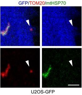
Vesicles containing selective mitochondrial cargo are found in proximity to the plasma membrane (≤1 µm) but away from the main mitochondrial network (>1 µm)
News Release, World Mitochondria Society, Berlin - Germany – September 2, 2022
Most cells constitutively secrete mitochondrial DNA and proteins in extracellular vesicles. While EVs are small vesicles that transfer material between cells, Mitochondria-Derived Vesicles (MDVs) carry material specifically between mitochondria and other organelles. Mitochondrial content can enhance inflammation under pro-inflammatory conditions, though its role in the absence of inflammation remains elusive.
- Germain et al. demonstrated that cells actively prevent the packaging of pro-inflammatory, oxidized mitochondrial proteins that would act as damage-associated molecular patterns (DAMPs) into EVs.
- They found that the distinction between material to be included into EVs and damaged mitochondrial content to be excluded is dependent on selective targeting to one of two distinct MDV pathways.
- They showed that Optic Atrophy 1 (OPA1) and sorting nexin 9 (Snx9)-dependent MDVs are required to target mitochondrial proteins to EVs, while the Parkinson’s disease-related protein Parkin blocks this process by directing damaged mitochondrial content to lysosomes.
These results provide insight into the interplay between mitochondrial quality control mechanisms and mitochondria-driven immune responses.
© Image -Todkar et al., Nature Communications 2022
Dr. Marc Germain will join Targeting Mitochondria 2022 to extensively discuss those result in a session entitled "Extracellular Vesicles & Mitochondria: The Target". Conference Program.
Media contact:
World Mitochondria Society
This email address is being protected from spambots. You need JavaScript enabled to view it.
+33-1-5504-7755
Targeting Mitochondria 2022 Congress
October 26-28, 2022 - Berlin, Germany
wms-site.com
More Articles...
- Transplantation of Astrocytic Mitochondria for Intracerebral Hemorrhage Treatment
- Researchers prove the potential of mitochondria-targeted chemotherapies
- Parkin in the Regulation of Myocardial Mitochondria-Associated Membranes and Cardiomyopathy During Endotoxemia
- Near-Infrared Light Modulation of Cytochrome C Oxidase in Traumatic Brain Injury


