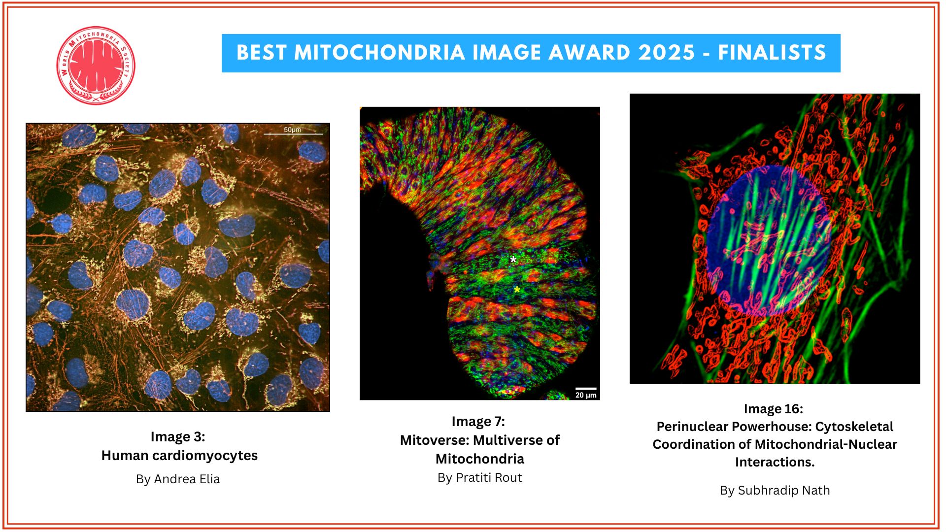
Nominations for Best Mitochondria Image 2024

1. Never Tear Us Apart
By Una Rastovic, The Roger Williams Institute of Liver Studies, King's College London & Foundation for Liver Research, London, UK
Study Context: The image depicts the mitochondrial network (TOM20, shown in orange) and Drp1 (in purple) within a proliferating hepatic stellate cell. Hepatic stellate cells are the primary producers of extracellular matrix (ECM) in liver fibrosis. During their activation in the fibrogenic process, these cells undergo proliferation, metabolic changes, and begin producing collagen along with other ECM proteins. This study seeks to investigate mitochondrial alterations during hepatic stellate cell activation, with the goal of developing mitochondria-targeted therapies for liver fibrosis.
2. Mitochondrial Networks and Communication between Cardiac Fibroblasts
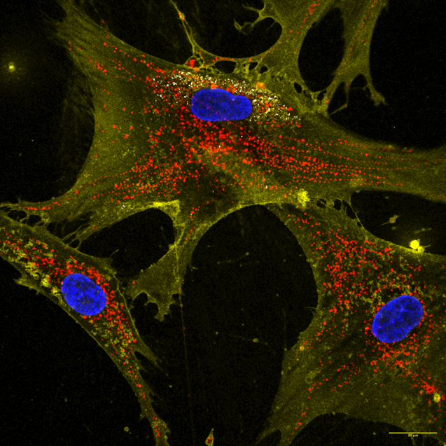
By Aybuke Celik, Research Fellow at Department of Cardiac Surgery in Boston Childrens Hospital/ Harvard Medical School
Study Context: This image reveals the complex mitochondrial network within cardiac fibroblasts (Mitotracker Red CMXRos). The central fibroblast extends its network toward neighboring cells, illustrating the dynamic intercellular communication essential for cellular energy metabolism and signal transduction. The image also highlights the cell membrane and cytoskeletal structures (Cell Mask Green), emphasizing the scaffolding that facilitates these cellular interactions. This visual insight underscores the pivotal role mitochondria play in cardiac fibroblast function, contributing to cardiac homeostasis and the maintenance of cellular signaling.
3. Mitochondrial network, a solar symphony and the nucleus, a lunar presence
By Nada Dhaouadi, PhD Student in the Histology laboratory at the polytechnic university of marches, under the supervision of Pr. Saverio Marchi
Study Description: In this captivating image, we observe the intricate architecture of mitochondria within a breast cancer cell, where the dynamic interplay between cellular structures creates a striking visual metaphor reminiscent of celestial bodies. The mitochondrial network, depicted in vibrant hues, radiates outward like the sun's rays, symbolizing energy production and vitality. Each mitochondrion appears as a luminous orb, interconnected in a web that suggests both strength and fragility. This network is not just a source of power for the cell; it embodies the relentless drive of cancer cells to proliferate and thrive, much like the sun's relentless energy that fuels life on Earth. In contrast, the nucleus stands out as a serene and powerful presence, akin to the moon illuminating the night sky. Its distinct shape and darker tones create a focal point within the chaotic beauty of the mitochondrial network. The nucleus houses the genetic material that dictates cellular function and behavior, representing both control and mystery—much like the moon's influence over tides and cycles. Together, the mitochondria and nucleus form a cosmic relationship within the breast cancer cell. The sun-like mitochondria provide energy and vitality, while the moon-like nucleus governs cellular processes with an enigmatic authority. This imagery not only highlights the complexity of cancer biology but also invites reflection on the dualities present in nature—light and dark, energy and control, chaos and order.
4. Mitochondria Movement in Neurons
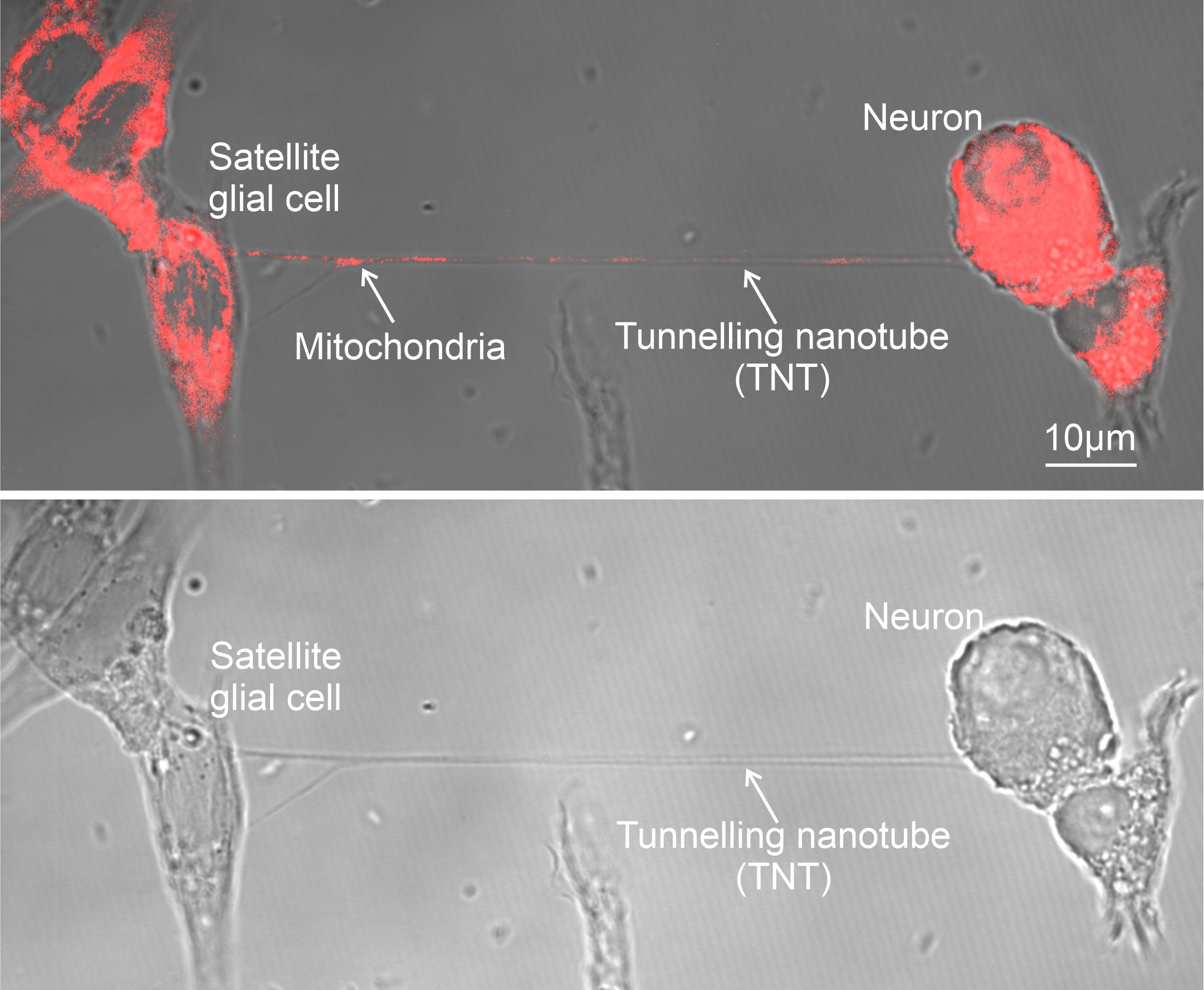
By Gaurav Verma, Department of Neuroscience and Physiology, Gothenburg University, Sweden
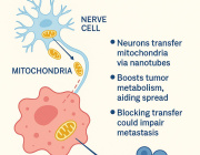
Study Description: Primary cortical neurons are grown in the coated (Poly-D-Lysin+Laminin) imaging plates and stained with MitoTracker Red, where we get a fascinating look at their energy production and overall health. MitoTracker Red is a special dye that stains active mitochondria. In neurons, this staining technique shows a complex network of mitochondria spread throughout the cell body, (axons and dendrites), highlighting where energy is being used the most. This allows us to track the movement of mitochondria movements from one neuron to others.
5. Mitochondrial Highways: Powering the Neuronal Network
By Amandine Grimm, Research Cluster Molecular and Cognitive Neurosciences, Department of Biomedicine, University of Basel, Switzerland
The image depicts a culture of induced pluripotent stem cells (iPSC)-derived neurons. The gray color indicates the nucleus (DAPI staining), the cyan color represents the neuronal marker MAP2 (microtubule-associated protein 2 staining), and the orange color represents the mitochondria (TOMM20 staining).
Study context: Our study aimed to investigate whether the mitochondrial aging signature is retained in iPSC-derived neurons from aged donors. IPSCs from both young and aged donors were differentiated into neurons before the assessment of mitochondria parameters. Notably, neurons from aged donors displayed mitochondrial dysfunction, including reduced ATP, mitochondrial membrane potential, and respiration, as well as increased free radicals and mitochondrial mass, and fragmented mitochondrial network compared to neurons from young donors. Our study indicates that aged iPSC-derived neurons could be a valuable tool for studying neuronal aging of mitochondrial parameters in vitro.
6. Forever young: Unravelling the lights of the eternal youth
By Vanessa López Polo, PhD student, Cellular Plasticity and Disease Laboratory, Institute for Research in Biomedicine (IRB), Barcelona, Spain
Senescent cell. Mitochondria was stained using mitotracker (magenta), mtDNA was stained using dsDNA antibody (green) and DNA was stained with DAPI (blue). Magnification: mtDNA staining resembles fireflies travelling along highly interconnected bridges which are mitochondria.
Study context: Senescent cells are used to study biological processes related to cellular aging and age-relate diseases. Senescent cells present an increase in the mitochondrial mass, increased in mtDNA number and hyperfused mitochondrial networks. Some cells present multiple nuclei.
7. Disruption of the Mitochondrial Network in Oxidative Stress-Induced Senescent Melanocytes
By Ines Martic, Institute for Biomedical Aging Research, Universität Innsbruck, Innsbruck, Austria
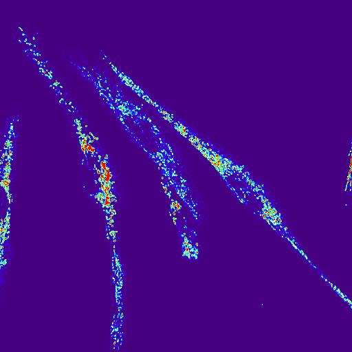
This complex V immunofluorescence image (60x magnification) in tBHP-treated melanocytes, highlights the mitochondrial network, with visible signs of increased fragmentation—an indicator of mitochondrial dysfunction associated with oxidative stress-induced senescence.
Study context: The study focuses on oxidative stress-induced senescence in melanocytes using tert-butyl hydroperoxide (tBHP) as a proxy for cigarette smoke exposure. Upon induction of senescence in melanocytes, the team aims to understand their role in skin aging and age-related pigmentation disorders. The observed mitochondrial fragmentation in tBHP-treated cells provides insight into how oxidative stress contributes to the cellular senescence, impairing mitochondrial function and promoting age-related phenotypes.
8. Parking Lot
By Dipti Baskar, National Institute of Mental Health and Neuro Sciences (NIMHANS), Bengaluru, India
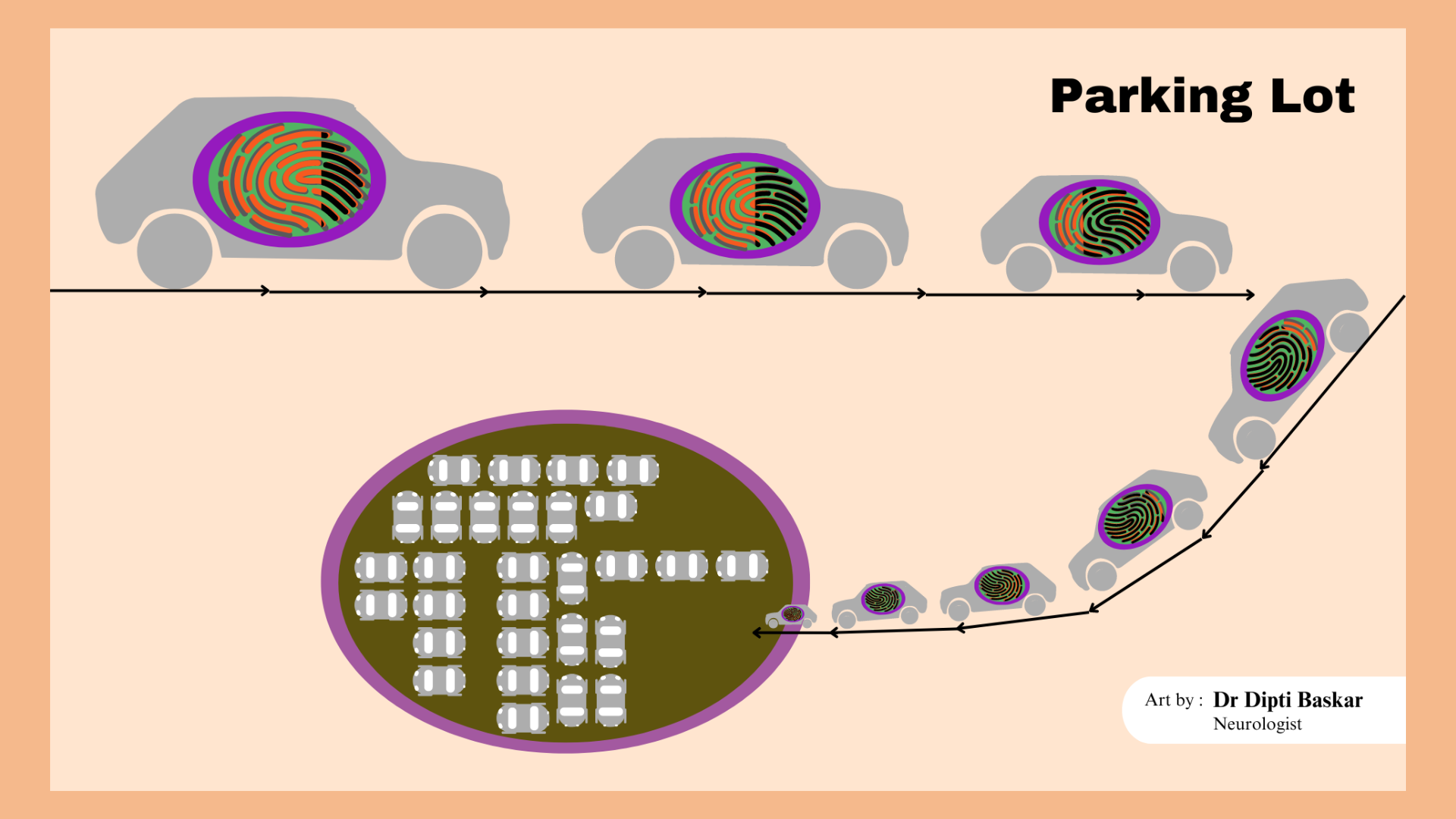
Description: Genetic abnormalities in Mitochondria, the "Power house" of the cell, result in "Power failure" of the body machinery. This can lead to paracrystalline inclusions called "Parking lot" inclusions within the abnormal mitochondria commonly noted in mitochondrial myopathy which is represented in this digital artwork.
Targeting Mitochondria 2024 Congress
October 29-31, 2024 - Berlin, Germany
wms-site.com


