


New research reveals a mitochondrial gene that protects against dementia and other diseases of aging
- Details
- Published on 28 September 2018
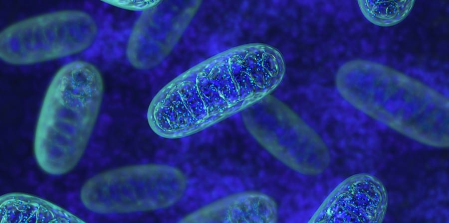
Image source: www.gero.usc.edu
New research from USC has uncovered a previously unknown genetic risk factor for Alzheimer’s disease and related dementias. The study provides insights on how these conditions, and other diseases of aging, might one day be treated and prevented.
The research from the Cohen Lab at the USC Leonard Davis School of Gerontology sheds new light on the protective role of a naturally occurring mitochondrial peptide, known as humanin. Amounts of the peptide decrease with age, leading researchers to believe that humanin levels play an important function in the aging process and the onset of diseases linked to older age.
“Because of the beneficial effects of humanin, a decrease in circulating levels could lead to an increase in several different diseases of aging, particularly in dementia,” said senior author Pinchas Cohen, dean of the USC Leonard Davis School and one of three researchers to independently discover the existence of humanin 15 years ago.
The study, Humanin Prevents Age-Related Cognitive Decline in Mice and is Associated with Improved Cognitive Age in Humans, led by Kelvin Yen of the USC Leonard Davis School, appears online on Sept. 21 in the Nature-published journal Scientific Reports. Among the findings, the researchers discovered a significant difference between the circulating levels of humanin in African-Americans, who are more impacted by Alzheimer’s disease and other diseases of aging, as compared to Caucasians.
“We have now discovered a novel underlying biological factor that may be contributing to this health disparity,” said Yen, a research assistant professor of gerontology.
Because humanin is encoded within the mitochondrial genome, the research team examined the mitochondrial DNA for common genetic variations known as SNPs (pronounced snips) that could explain the differences between humanin levels. According to the paper, after sequencing the entire mitochondrial genome of all samples to determine if any SNPs correlated with humanin levels, they identified a genetic variation that was associated with a 14 percent decrease in circulating humanin levels.
This provides the first evidence that a variation in the sequence of a mitochondrial peptide is associated with a change in the level of peptides and the first conclusive demonstration that mitochondrial peptides are encoded in and regulated through mitochondrial DNA, Cohen said.
The team subsequently examined the effect of this SNP in a separate large cohort of participants from the Health and Retirement Study, a longitudinal study of approximately 20,000 individuals over the age of 50 in the United States, of which more than 12,500 individuals consented to DNA analysis. Using this data set, the researchers report finding that the unfavorable version of this SNP, associated with low levels of the humanin peptide, is also associated with accelerated cognitive aging, providing the first demonstration linking a humanin SNP in the mitochondria to cognitive decline in people.
The paper also shows that in mice, injections of humanin delay cognitive decline associated with aging, proposing a possible therapeutic role for humanin-related drugs. The mechanism through which humanin acts to mediate these effects involves suppression of inflammation systemically, as well as specifically in the brain.
News source: www.gero.usc.edu
Mitochondrial diseases could be treated with gene therapy
- Details
- Published on 28 September 2018
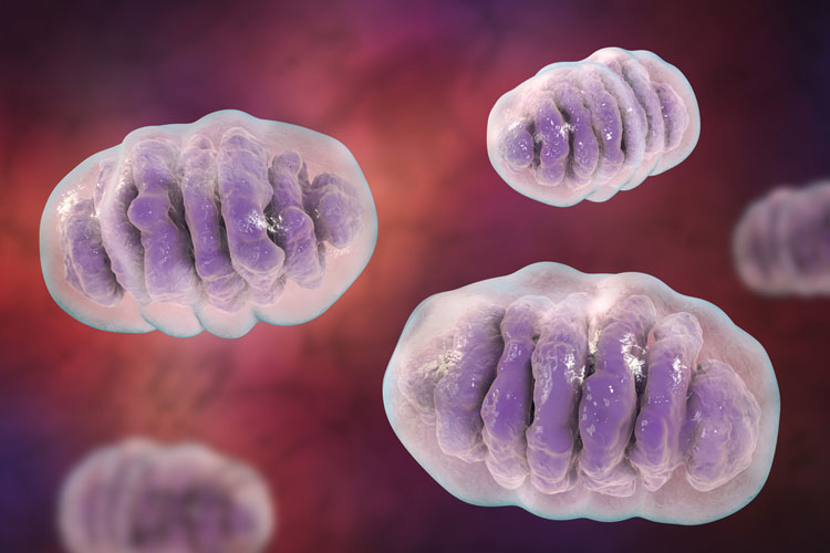
Image source: drugtargetreview.com
Researchers have developed a genome-editing tool for the potential treatment of mitochondrial diseases: serious and often fatal conditions which affect 1 in 5,000 people.
The researchers, led by the University of Cambridge, applied an experimental gene therapy treatment in mice and were able to successfully target and eliminate the damaged DNA in mitochondria which causes the devastating conditions.
Their results could provide a practical route to treating patients with these diseases and may provide a future alternative to mitochondrial replacement therapy, or ‘three-parent IVF’. This is the first time programmable genome engineering tools have been used inside a living animal, resulting in such significant modification of mitochondrial DNA.
Mitochondria are the powerhouses inside our cells, producing energy and carrying their own DNA. They are inherited from a person’s mother via the egg, but if they are damaged, it can result in a serious mitochondrial disease. For example, MELAS Syndrome is a severe multi-system disorder causing progressive loss of mental and movement abilities, which usually becomes apparent in early childhood.
There are typically about 1000 copies of mitochondrial DNA per cell, and the percentage of these that are damaged, or mutated, will determine whether a person will suffer from mitochondrial disease or not. Usually, more than 60% of the mitochondrial DNA molecules in a cell need to be mutated for the disease to emerge, and the more mutated mitochondrial DNA a person has, the more severe their disease will be. Conversely, if the percentage of mutated DNA can be reduced, the disease could potentially be treated.
Mitochondrial diseases are currently incurable, although a new IVF technique of mitochondrial transfer gives families affected by mitochondrial disease the chance of having healthy children – removing affected mitochondria from an egg or embryo and replacing them with healthy ones from a donor.
“Mitochondrial replacement therapy is a promising approach to prevent transmission of mitochondrial diseases, however, as the vast majority of mitochondrial diseases have no family history, this approach might not actually reduce the proportion of mitochondrial disease in the population,” said Dr Payam Gammage, a postdoctoral researcher in the MRC Mitochondrial Biology Unit, and the paper’s first author.
“One idea for treating these devastating diseases is to reduce the amount of mutated mitochondrial DNA by selectively destroying the mutated DNA, and allowing healthy DNA to take its place,” said Dr Michal Minczuk, also from the Medical Research Council (MRC) Mitochondrial Biology Unit, and the study’s senior author.
To test an experimental gene therapy treatment, which has so far only been tested in human cells grown in petri dishes in a lab, the researchers used a mouse model of mitochondrial disease that has the same mutation as some human patients.
The gene therapy treatment, known as the mitochondrially targeted zinc finger-nuclease, or mtZFN, recognises and then eliminates the mutant mitochondrial DNA, based on the DNA sequence differences between healthy and mutant mitochondrial DNA. As cells generally maintain a stable number of mitochondrial DNA copies, the mutated copies that are eliminated are replaced with healthy copies, leading to a decrease in the mitochondrial mutation burden that results in improved mitochondrial function.
The treatment was delivered into the bloodstream of the mouse using a modified virus, which is then mostly taken up by heart cells. The researchers found that the treatment specifically eliminates the mutated mitochondrial DNA, and resulted in measures of heart metabolism improving.
Following on from these results, the researchers hope to take this gene therapy approach through clinical trials, in the hope of producing an effective treatment for mitochondrial diseases.
This work was supported by the Medical Research Council and was performed in collaboration with Sangamo Therapeutics and the Max Planck Institute for Biology of Ageing in Cologne.
News source: www.drugtargetreview.com
Reference: Payam A. Gammage et al. ‘Genome editing in mitochondria corrects a pathogenic mtDNA mutation in vivo.’ Nature Medicine (2018). DOI: 10.1038/s41591-018-0165-9.
Study Provides New Insights For Ways to Use Cell Metabolism to Treat Cancer
- Details
- Published on 31 August 2018

Researchers at the University of Cincinnati (UC) College of Medicine have discovered that cell metabolism plays an important role in the ability of cells to start a survival program called autophagy, an unwanted side effect of some anti-cancer drugs that helps some tumor cells dodge treatment and eventually regrow into new tumors.
These findings, reported in the Aug. 28 online edition of the journal Cell Reports, provide new insights for ways to use cell metabolism to "pull the plug" on tumor cells that survive treatment, possibly leading to better treatments and outcomes for patients.
"Cells adapt to nutrient starvation by increasing autophagy, where a cell basically eats itself and recycles cellular contents to support essential processes until nutrients become plentiful once again. This process is regulated by the mammalian target of rapamycin (mTOR) and AMP-activated protein kinases (AMPK)," says Carol Mercer, Ph.D., research assistant professor in the Division of Hematology Oncology, Department of Internal Medicine, and a member of both the Cincinnati Cancer Center and UC Cancer Institute. "Drugs that target mTOR or activate AMPK are being used in the clinic for some cancers, and are under active investigation for others, making it important to understand how they affect this tumor cell survival pathway."
"We found that cell metabolism significantly influences the ability to begin autophagy, with mitochondrial complex I function being an important factor in the initiation, amplification and duration of the response," she continues. "We show that the anti-diabetic drug phenformin, the anti-diabetic drug metformin, and genetic defects in complex I shift cell metabolism toward glycolysis and inhibit the ability of mTOR inhibitors to prompt autophagy. The opposite is also true, as a shift away from glycolysis and toward mitochondrial metabolism, enhances autophagy through a mechanism that involves increased phospholipid metabolism. Our data demonstrate the importance of metabolism in the regulation of autophagy, increase our understanding of clinically relevant drugs that are important for cancer, and suggest new strategies to increase or inhibit autophagy."
Mercer, principal investigator on the study, and her lab, worked primarily in cultured cells to understand how metabolism regulates autophagy, identifying strategies to manipulate this pathway to the patients' advantage. This work was built on pre-clinical studies in animal models by Hala Elnakat Thomas, Ph.D., first author and research instructor in the department, who found that the combination of mTOR inhibitors were effective in the treatment of hepatocellular carcinoma (liver cancer) but had the potential disadvantage of increasing autophagy.
"Our data reveal the dynamic and metabolic regulation of autophagy and suggest new therapeutic strategies for cancer, neurodegenerative and mitochondrial diseases," Mercer says. "We need to further explore the reasons this occurs and the implications for how the metabolic regulation of autophagy can be used in the clinic."
News source: www.rdmag.com
More information: Mitochondrial Complex I Activity Is Required for Maximal Autophagy , Cell Reports (2018). DOI: 10.1016/j.celrep.2018.07.101
https://www.cell.com/cell-reports/fulltext/S2211-1247
Liver disease drug could help restore cells damaged by Alzheimer's
- Details
- Published on 05 September 2018
A drug which has been used to treat liver disease for decades could help to restore cells damaged by Alzheimer's, a new study from the University of Sheffield has found.
The pioneering study, funded by Alzheimer's Research UK, discovered the drug ursodeoxycholic acid (UDCA) improves mitochondrial dysfunction – which is known to be a causative factor for both sporadic and familial Alzheimer's disease.
Mitochondria play a pivotal role in both neuronal cell survival and death as they regulate energy metabolism and cell death pathways acting as a cell's battery.
Mitochondrial abnormalities have been identified in many cell types in Alzheimer's disease, with deficits occurring before the development of the classical pathological aggregations. The energy changes have been found in many different cells from people with Alzheimer's. It is thought they are one of the earliest changes to occur in the brain cells, perhaps even before symptoms are reported by people living with the disease.
Dr Heather Mortiboys, Parkinson's UK Senior Research Fellow at the University of Sheffield's Institute of Translational Neuroscience (SITraN), said: "For the first time in actual Alzheimer's patient tissue this study has shown that the drug UDCA acid can boost the performance of the cells' batteries, the mitochondria.
"We also found that the drug, which is already in clinical use for liver disease, acts by changing the shape of the batteries which could tell us more about how other drugs can be beneficial in Alzheimer's.
"Most importantly we found the drug to be active in cells from people with the most common type of the devastating disease – sporadic Alzheimer's – which could mean it has potential for thousands of patients."
Dr Mortiboys, who led the study, added: "As the drug is already in clinical use for liver disease; this speeds up the potential time it could take to get this drug to the clinic for patients."
The ground-breaking research also found the drug changed the shape of mitochondria by redistributing Dynamin-related protein 1 (Drp1) to the mitochondria in people with Alzheimer's skin cells. Drp1 is a regulator of mitochondrial shape and locates at the mitochondria to initiate fission events. It is thought this could have neuroprotective effects in Alzheimer's disease. This study suggests this pathway could be manipulated by drugs which are then neuroprotective in patients themselves.
The next steps could include studies in patient-derived neurons to check for protective effects or, as others have already shown UDCA to be protective in animal models of Alzheimer's disease, steps could be taken to move UDCA to clinical trials.
Alzheimer's disease is the leading cause of dementia worldwide and is the most common neurodegenerative disorder. It currently affects 850,000 people in the UK, with numbers expected to soar to two million by 2051.
Dr Sara Imarisio, Head of Research at Alzheimer's Research UK, who funded the work, said:"Today, around half a million people in the UK are living with Alzheimer's disease. With no new dementia drugs in over 15 years, it's vital we continue to approach Alzheimer's from as many angles as possible.
"Through innovative research we are building a clearer picture of the complexities of the disease and how it develops in the brain. This work suggests a potential new way to target Alzheimer's but needs further exploration before we can know whether this drug used for a liver condition is safe or effective for people with Alzheimer's disease.
"Alzheimer's Research UK receives no government funding for the research we support, and it is only thanks to the generosity of our supporters that we're able to fund vital projects like this."
Previous laboratory studies conducted by SITraN in 2015 showed UDCA could be an effective treatment in halting the progression of Parkinson's disease. The collaborative study demonstrated the effects of the drug in patients that carry the LRRK2 mutation. The study showed improved mitochondrial function as demonstrated by the increase in oxygen consumption and cellular energy levels.
The new research is published in the Journal of Molecular Biology.
News source: www.medicalxpress.com
More information: Shilpy Dixit et al. Mitochondrial dysfunction in the APP/PSEN1 mouse model of Alzheimer's disease and a novel protective role for ascorbate, Free Radical Biology and Medicine (2017). DOI: 10.1016/j.freeradbiomed.2017.08.021
Scientists reverse Aging-associated skin wrinkles and hair loss in a mouse model
- Details
- Published on 23 July 2018

Wrinkled skin and hair loss are hallmarks of aging. What if they could be reversed?
Keshav Singh, Ph.D., and colleagues have done just that, in a mouse model developed at the University of Alabama at Birmingham. When a mutation leading to mitochondrial dysfunction is induced, the mouse develops wrinkled skin and extensive, visible hair loss in a matter of weeks. When the mitochondrial function is restored by turning off the gene responsible for mitochondrial dysfunction, the mouse returns to smooth skin and thick fur, indistinguishable from a healthy mouse of the same age.
"To our knowledge, this observation is unprecedented," said Singh, a professor of genetics in the UAB School of Medicine.
Importantly, the mutation that does this is in a nuclear gene affecting mitochondrial function, the tiny organelles known as the powerhouses of the cells. Numerous mitochondria in cells produce 90 percent of the chemical energy cells need to survive.
In humans, a decline in mitochondrial function is seen during aging, and mitochondrial dysfunction can drive age-related diseases. A depletion of the DNA in mitochondria is also implicated in human mitochondrial diseases, cardiovascular disease, diabetes, age-associated neurological disorders and cancer.
"This mouse model," Singh said, "should provide an unprecedented opportunity for the development of preventive and therapeutic drug development strategies to augment the mitochondrial functions for the treatment of aging-associated skin and hair pathology and other human diseases in which mitochondrial dysfunction plays a significant role."
The mutation in the mouse model is induced when the antibiotic doxycycline is added to the food or drinking water. This causes depletion of mitochondrial DNA because the enzyme to replicate the DNA becomes inactive.
In four weeks, the mice showed gray hair, reduced hair density, hair loss, slowed movements and lethargy, changes that are reminiscent of natural aging. Wrinkled skin was seen four to eight weeks after induction of the mutation, and females had more severe skin wrinkles than males.
Dramatically, this hair loss and wrinkled skin could be reversed by turning off the mutation. The photos below show the hair loss and wrinkled skin after two months of doxycycline induction, and the same mouse a month later after doxycycline was stopped, allowing restoration of the depleted mitochondrial DNA.
Little change was seen in other organs when the mutation was induced, suggesting an important role for mitochondria in skin compared to other tissues.
The wrinkled skin showed changes similar to those seen in both intrinsic and extrinsic aging -- intrinsic aging is the natural process of aging, and extrinsic aging is the effect of external factors that influence aging, such as skin wrinkles that develop from excess sun or long-term smoking.
Among the details, the skin of induced-mutation mice showed increased numbers of skin cells, abnormal thickening of the outer layer, dysfunctional hair follicles and increased inflammation that appeared to contribute to skin pathology. These are similar to extrinsic aging of the skin in humans. The mice with depleted mitochondrial DNA also showed changed expression of four aging-associated markers in cells, similar to intrinsic aging.
The skin also showed disruption in the balance between matrix metalloproteinase enzymes and their tissue-specific inhibitor -- a balance of these two is necessary to maintain the collagen fibers in the skin that prevent wrinkling.
The mitochondria of induced-mutation mice had reduced mitochondrial DNA content, altered mitochondrial gene expression, and instability of the large complexes in mitochondria that are involved in oxidative phosphorylation.
Reversal of the mutation restored mitochondrial function, as well as the skin and hair pathology. This showed that mitochondria are reversible regulators of skin aging and loss of hair, an observation that Singh calls "surprising."
"It suggests that epigenetic mechanisms underlying mitochondria-to-nucleus cross-talk must play an important role in the restoration of normal skin and hair phenotype," Singh said, who has a secondary UAB appointment as professor of pathology. "Further experiments are required to determine whether phenotypic changes in other organs can also be reversed to wildtype level by restoration of mitrochondrial DNA."
News source: https://www.rdmag.com
Singh B, Schoeb TR, Bajpai P, Slominski A, Singh KK. Reversing wrinkled skin and hair loss in mice by restoring mitochondrial function.
Cell Death Dis. 2018, 9:735, DOI: 10.1038/s41419-018-0765-9


