Best Image Award


- Details
- Published on 27 May 2025

Best Scientific Image Award 2026: Show the World Your Mitochondrial Discovery
Can a single image change the way we see mitochondrial biology?
The World Mitochondria Society (WMS) is pleased to announce the opening of submissions for the Best Scientific Image Award 2026, an international competition celebrating the most outstanding scientific images in mitochondrial research.
From stunning microscopy to innovative scientific illustrations and AI-assisted visualizations, this award recognizes images that combine scientific excellence with visual impact. Every image tells a story, reveals a discovery, and helps communicate the remarkable complexity of mitochondria.
The winner will be announced during the Awards Ceremony of the 17th World Congress on Targeting Mitochondria, to be held in Berlin, Germany, October 21–23, 2026.
Who can participate?
The competition is open to researchers, clinicians, postdoctoral fellows, PhD students, engineers, and imaging specialists from academia and industry worldwide.
What can you submit?
We welcome original scientific images related to mitochondrial biology, including:
- Fluorescence and confocal microscopy
- Electron microscopy
- Live-cell imaging
- Super-resolution microscopy
- Clinical and translational imaging
- Scientific illustrations
- AI-assisted scientific visualizations
How to submit:
Each submission should include a high-resolution image, a title, a short scientific description (maximum 250 words), and the imaging technique used.
mitochondria[at]wms-site.com
Awards
The Best Scientific Image Award 2026 winner will receive:
- An official World Mitochondria Society Award Certificate
- Free registration to the 17th World Congress on Targeting Mitochondria 2026
- Recognition during the Awards Ceremony in Berlin
- International visibility through the WMS website, newsletter, and social media
- Inclusion in the WMS Scientific Image Gallery
New for 2026: People's Choice Award
For the first time, congress participants will vote for their favorite image during the meeting. This special award will recognize the image that best inspires and communicates mitochondrial science to the international community.
Submit Your Image
Submission deadline: September 20, 2026
Whether your work explores mitochondrial dynamics, metabolism, signaling, aging, disease, or therapeutic innovation, we encourage you to share the image that best represents your science.
One image can inspire a discovery. One image can inspire a community. We look forward to seeing yours.

We are thrilled to reveal the three winners of the Best Image in Mitochondria Research 2025!
Their exceptional visuals captured the beauty, complexity, and dynamism of mitochondrial science, merging art and discovery.
Winners (ex aequo):
1️ “Human Cardiomyocytes” – Andrea Elia
2️ “Mitoverse” – Pratiti Rout
3️ “Perinuclear Powerhouse” – Subhradip Nath
Each winner receives:
Free registration for Targeting Mitochondria 2025 or 2026
Guaranteed Short Oral Presentation slot during the 2026 Congress
Congratulations to our talented winners — and thank you to all participants who helped make this edition a visual celebration of mitochondrial research.
Together, we continue to show that mitochondria are not only the powerhouse of the cell, but also a source of inspiration and innovation.
Discover more highlights from Targeting Mitochondria 2025 – Berlin
All Submissions received
Image 1: Lighting Up Mitochondrial Stress: SNX9 Dynamics in Parkinson’s disease
By Jimna Mohamed Ameer, PhD student, Inter University Centre for Biomedical Research and Super Specialty Hospital, Mahatma Gandhi University, Kottayam, Kerala, India
Description : A Confocal image of an MPP⁺, mitochondrial complex inhibitor-treated differentiated SH-SY5Y cell showing recruitment of SNX9 protein to mitochondria. Mitochondria were stained with MitoTracker (red), SNX9 was detected using a specific antibody (green), and nuclear DNA was counterstained with DAPI (blue).
Context of the Study : MPP⁺ -induced mitochondrial dysfunction in differentiated SH-SY5Y cells models early events in Parkinson’s disease. This neurotoxic stress causes mitochondrial fragmentation and disrupts endolysosomal functions. Our results reveal that Sorting Nexin 9 (SNX9), a membrane remodelling protein involved in endocytosis, is recruited to dysfunctional mitochondria following MPP⁺ treatment. This suggests a potential role for SNX9 in mitochondrial-derived vesicle (MDV) formation, providing novel insights into mitochondrial signalling and vesicular trafficking during neurodegenerative stress responses.
Image 2: Glow and behold: Mitochondria on the move
By Phua Qian Hua, Institute of Molecular and Cell Biology, Agency for Science, Technology and Research (ASTAR), Singapore, National University of Singapore
Description: This image captures our mitochondrial transplantation experiment, where healthy isogenic mitochondria were isolated from iPSCs and transplanted into MELAS endothelial cells, characterized by mitochondrial dysfunction. Confocal microscopy image taken after mitochondrial transplantation from healthy iPSC-derived mitochondria (labeled with MitoTracker Green) into diseased endothelial cells with dysfunctional endogenous mitochondria (labeled with MitoTracker Red).
Study Context: This experiment aimed to assess mitochondrial uptake and evaluate whether transplantation could restore mitochondrial and endothelial cell function.
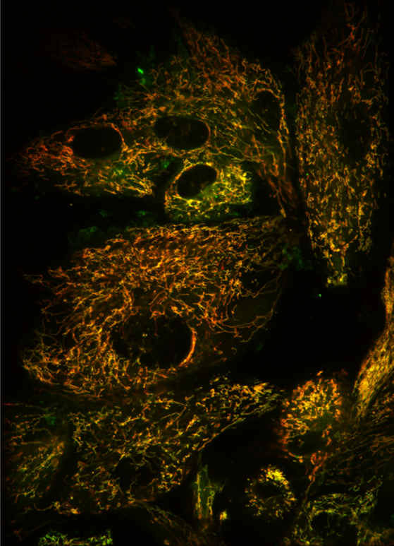
Image 3: Human cardiomyocytes
By Andrea Elia, Alzheimer's Center at Temple (ACT), Department of Neural Sciences, Lewis Katz School of Medicine, Temple University, Philadelphia
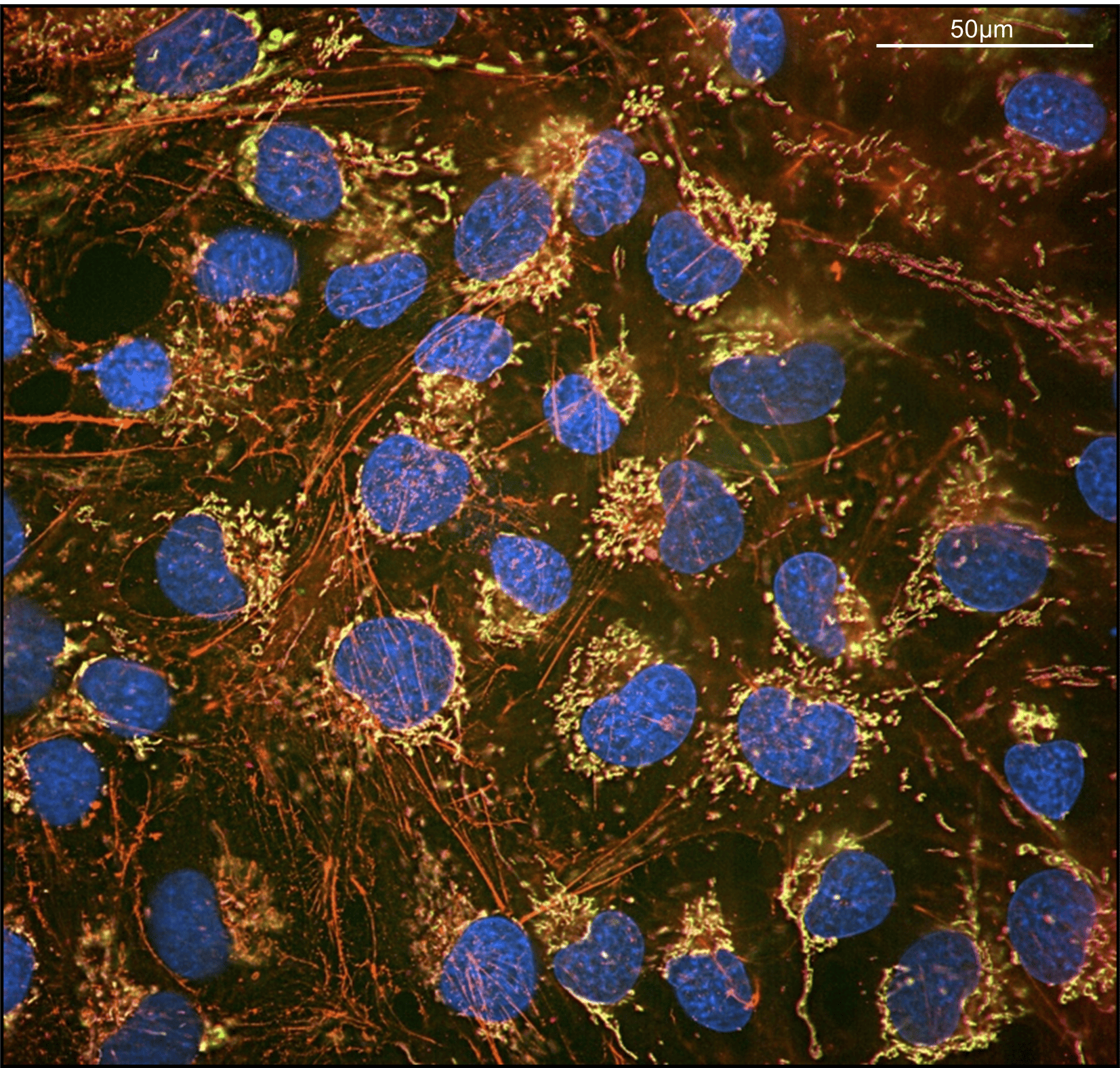
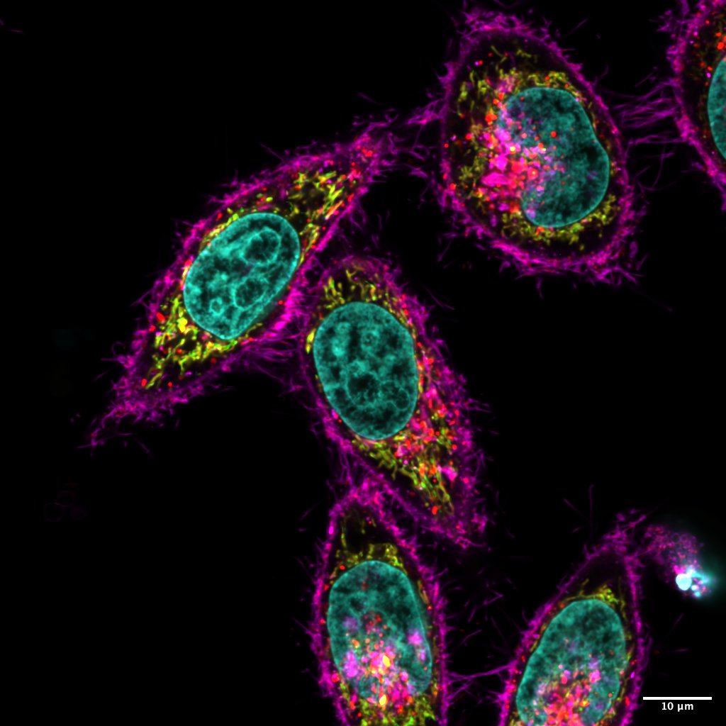
Description : Human Aβ40 oligomers disrupt mitochondrial architecture and trigger cytochrome C release in human cardiomyocytes (AC16). Representative digital images (scale bar 50μm) of AC16, stained with TOM20 (magenta), cytochrome c (CytC, green), Phalloidin (red), and DAPI (blue).
Study context: Alzheimer’s disease (AD) is a multifaceted neurodegenerative disorder primarily defined by the accumulation of cerebral amyloid-β (Aβ) plaques and tau protein tangles. Notably, approximately 90% of AD patients also develop cerebral amyloid angiopathy (CAA), a condition marked by Aβ deposits within and surrounding cerebral blood vessels, which exacerbates vascular damage, neuroinflammation, blood-brain barrier (BBB) disruption, and neuronal loss. Among the vascular mechanisms implicated in AD progression, dysfunction in cerebrovascular mitochondrial metabolism emerges as a key driver of Aβ-induced cerebral endothelial cell (cECs) apoptosis, heightened BBB permeability, and inflammation. Our previous studies demonstrated that the vasculotropic Aβ40 variant, strongly associated with CAA, triggers cECs' death via mitochondrial impairment and cytochrome c (CytC) release, both in vitro and in vivo. Increased BBB permeability in AD may facilitate systemic dissemination of Aβ40, promoting its accumulation in peripheral organs such as the heart. Yet, the impact of Aβ on cardiac cell homeostasis and function remains largely unexplored. In this study, we investigated cardiac physiology and amyloid pathology in the hearts of Tg2576-AD-CAA mice and examined the mitochondrial mechanisms through which Aβ40 affects human cardiomyocytes. Our findings establish human cardiomyocytes as a robust model for uncovering the pathological effects of AD-associated Aβ on cardiac function. Specifically, we show that Aβ40 oligomers compromise mitochondrial structure and destabilize cytochrome c in cardiomyocytes, suggesting a novel pathway through which Aβ contributes to cardiac dysfunction in AD.
Image 4: Glowing and Growing Mind: Mitochondria in Early Neural Development
By Sundas Arshad, Luxembourg centre for Systems Biomedicine, University of Luxembourg
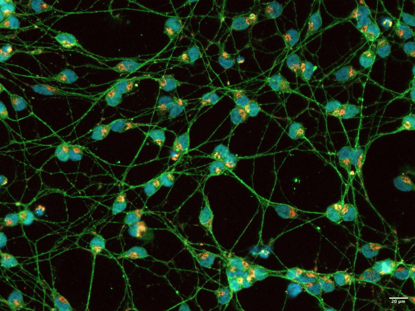
Description : This fluorescent image represents LUHMES-derived dopaminergic neurons on Day 3 of differentiation. Nuclei are marked with DAPI (blue), TH positive dopaminergic neurons (Green), and mitochondria are stained with TOM20 (red), illuminating their vibrant distribution along the delicate neuronal processes. The dynamic mitochondrial network signifies the early stages of neuronal development and energy demand as these cells begin to specialize.
Study Context : This image is part of a study investigating mitochondrial remodeling during early neuronal differentiation. LUHMES cells, which are human embryonic neuronal precursor cells, have been utilized in several studies to investigate the PD and underlying mechanisms associated with it. Mitochondria were stained using TOM20, a marker of the outer mitochondrial membrane, to visualize their distribution and morphology. The goal of our study is to investigate the crosstalk between Ca²⁺ signaling and mitochondrial dynamics in dopaminergic neurons by combining live cell imaging and immunostaining techniques.
Image 5: Bumpy mitochondrial track: a roadway to disease
By Ritoprova Sen, Integrated Ph.D. student of Biological Sciences, Jawaharlal Nehru Centre for Advanced Scientific Research, Bengaluru, India
Description: This is a projected fluorescent microscopy image depicting an AC16 human cardiomyocyte cell expressing a dominant negative disease mutant exhibiting hyper-elongated mitochondria. The outer mitochondrial membrane has been labelled with a construct expressing OMP25 (labelled in magenta) while the green denotes COXIV, an inner mitochondrial membrane protein which is a part of complex IV of the electron transport chain. The mutant manifests as elongated mitochondria, interrupted by bumps in the form of swollen bulbs as seen in magenta. Interestingly these swollen bulbs do not costain with COXIV in green. Whether such mitobulbs cause localized mitochondrial damage and consequently activation of mitochondrial quality control is currently an active area of investigation in the lab.
Study Context : Our study aims to investigate mitochondrial damage and quality control in the context of mitochondrial diseases, particularly in cardiomyocytes. One such mitochondrial disease associated mutant causes elongated mitochondria as observed in AC16 human cardiomyocyte cell line.
Image 6: Galacto’Mitochondria in Cancer universe
By Lea Di Mascio, Research Engineer in molecular biology, Team 3 “ Metabolism, Cancer and Immune Response” at the Molecular Medicine Mediterranean Center (C3M) of Nice
Description: Here you can observe in A549 cells (Human cell line) the interaction between 2 mitochondrial proteins, one localized in the outer membrane of mitochondria (TOM20) and the other only visible under stress condition (PINK1). This experiment allows us to be sure that mitophagy is involved in lung cancer. TOM20 is red. Because we are under stress, the mitochondrial network is completely fragmented. PINK1 is green and only stabilized under stress condition. The colocalization between these two proteins is orange. This image is really interesting because we can easily observe the 3 colors and the explosion of the mitochondrial network in lung cancer like galaxies in universe.
Context of Study: As mitophagy is a major process that allows cancer cells to resist both environmental stress and chemotherapeutic agents and because lung cancer highly relies on mitochondrial metabolism, our team focuses on the mitochondria turn over during lung cancer progression. We are following the mitochondria metabolism at early and late stage of lung cancer progression (in vivo and in vitro) and we try to understand how the mitophagy is involved in the development and resistance to treatment.
Image 7: Mitoverse: Multiverse of Mitochondria
By Pratiti Rout, PhD Scholar Justus-Liebig-Universität Gießen and doing part of her doctoral research at Philipps-Universität Marburg
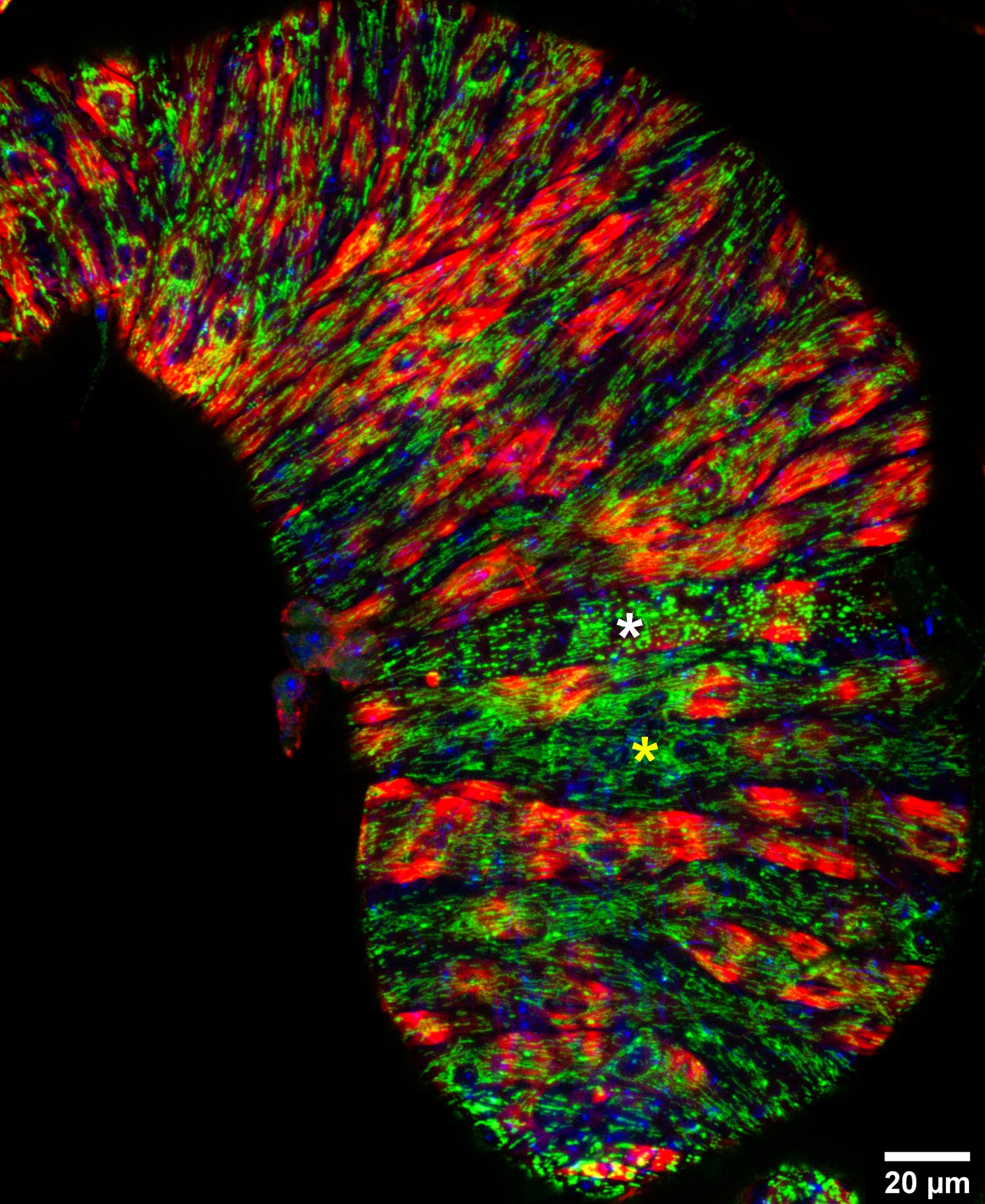
Description: The image showcases how in a single tissue (seminal vesicle of Drosophila male reproductive system) mitochondria can adapt to such diverse architectures (interconnected networks- * in yellow, fragmented and globular shapes - * in white). Phalloidin marks actin in red, DAPI stains the nuclei in blue and mitochondria in the muscles of seminal vesicle is visualised in green with Mef2-Gal4 driven UAS-mitoGFP. MEF is muscle-specific transcription factor.
Study Context: Mitochondrial dynamics is crucial for muscle function. In this study, we explore the role of mitochondrial architecture in muscle development and function using Drosophila as a model organism. In our attempt to observe differences in mitochondria morphology in different muscle types found in Drosophila male reproductive tissues, we aquired this image using Leica Stellaris confocal microscope. Accessory glands like seminal vesicles are usually surrounded by multinucleated striated muscles whereas testes has smooth-like muscles. Surprisingly, we observed different shapes of mitochondria in muscles of one single tissue (seminal vesicle) compelling us to reflect on the idea of 'multiverse of mitochondria'.
Image 8: A Quiet Outlier : Mitochondrial Harmony in a Fragmented Cellscape
By Doğa Buse Köm, MSc Student, Graduate School of Health and Sciences, Koc University, Istanbul, Turkey
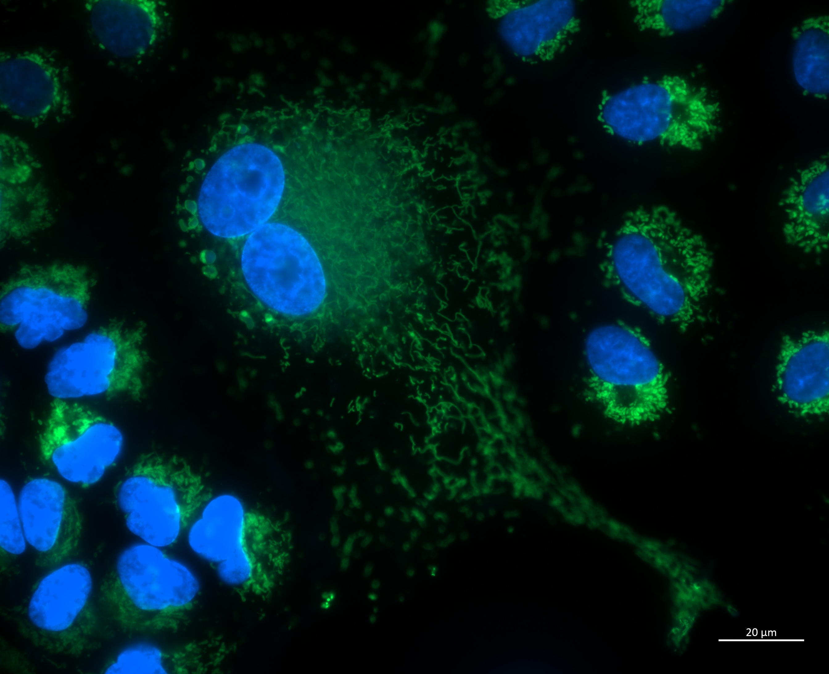
Description : The mitochondrial architecture showed remarkable cell-to-cell diversity, as shown by fluorescence imaging. The majority of cells have fragmented morphologies and a compact, perinuclear distribution of mitochondria. A large, highly reticulated mitochondrial network, on the other hand, is visible in a prominent central cell, indicating a fused and metabolically active state. This unique shape could be a result of variations in the surrounding microenvironment, mitochondrial health, or cell cycle phase.
Study Context: Hoechst 33342 and MitoTracker Green were used to stain A549 cells to see the morphology of the nucleus and mitochondria. Understanding how cellular context and culture conditions affect mitochondrial dynamics and how these alterations relate to structural and functional modifications in epithelial cancer cells is the goal of the study.
Image 9 - Mito-art : Stopping to enjoy the process
By Tania Arguello Saenz, Ph.D, Carlos Moraes Laboratory, University of Miami, Fl, USA
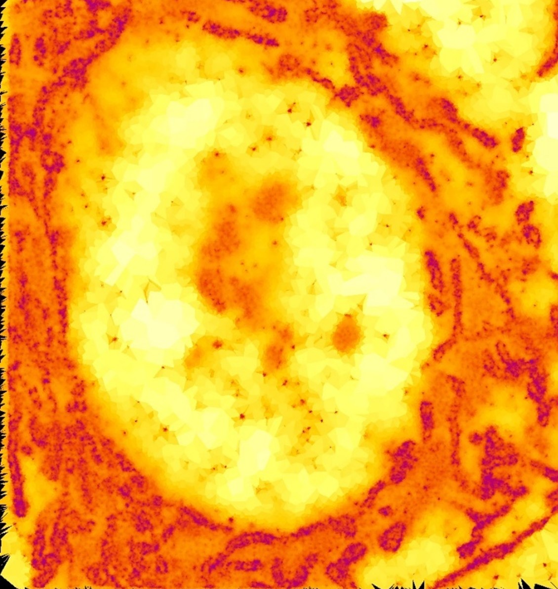
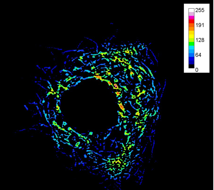
Description: Localizing events of the mitochondrial outer membrane protein TOM20 antibody signal surrounding the nucleus in a HEK293T cell using gradient color, Voronoi tessellation filling polygons (512 x 512, 10,000 frames)"
Study Context: Single-molecule localization microscopy (SMLM) process analysis of the mitochondrial outer membrane protein TOM20 used as a reference to measure colocalization events with other not known (not shown) outer and/or inner mitochondrial membrane proteins.
Image 10: The Green Thread of Life
By Sambeet Jena, BS-MS at Neuroscience and Stem Cell Biology Lab, School of Biological Sciences, National Institute of Science Education and Research, Bhubaneswar, India
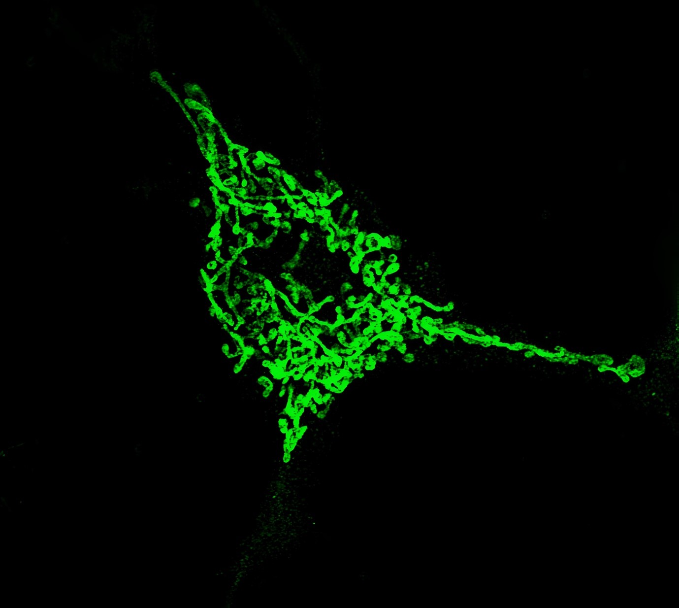
Subtitle: Super-resolution imaging of TOMM20-labelled mitochondria reveals the elegant architecture of the cell’s energy network.
Description: This image captures mitochondria in striking detail using ELYRA SIM super-resolution microscopy. Labelled with TOMM20 to mark the outer mitochondrial membrane, these glowing green filaments weave through the cytoplasm, connecting sites of energy demand with the source that powers them. In neurons, these threads are more than structural, they are essential for memory, survival, and synaptic plasticity. Here, they stand as the silent green threads holding life together.
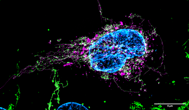
Image 11 - Inflamed Harmony : Red Echoes Around a Blue Core
By Jitesh Gupta,Dept. of Animal Biology, School of Life Sciences, University of Hyderabad, India

Description : Confocal image of a human corneal epithelial cell showing an interconnected mitochondrial network (red) surrounding the nucleus (blue) under inflammatory stress.
The image shows a fluorescence micrograph of a human corneal epithelial cell treated with interferon (IFN) and tumour necrosis factor (TNF), illustrating mitochondrial morphology under inflammatory conditions. The nucleus is prominently stained blue with a DNA-binding dye (such as DAPI) and appears intact and centrally located. Mitochondria, labelled in red, display a fragmented and punctate distribution around the nucleus, deviating from the typical elongated, interconnected network observed in healthy cells. This mitochondrial fragmentation is indicative of inflammation-induced stress and is commonly associated with activation of mitochondrial fission pathways, disrupted mitochondrial dynamics, and potential initiation of apoptotic signaling. The altered morphology reflects the impact of IFN and TNF on mitochondrial integrity and function in human corneal epithelial cells, highlighting the role of mitochondrial remodelling during inflammatory responses in ocular surface tissues.
Image 12 - A New Light on Mitochondria: Multiplexed Visualization in Living Cells
By Natalija Trunkelj, Msc Pharmacy, 1st year PhD student, University of Ljubljana, Faculty of Phamracy, Slovenia
Description: HeLa cells stained using our newly developed fluorescent mitochondrial dye (excited at 405 nm), revealing mitochondrial structures with high specificity. The nuclei are stained with Hoechst 33342 (excited at 405 nm), lysosomes with LysoTracker™ Red DND-99 (excited at 561 nm), the cell membrane with CellMask™ Deep Red (excited at 640 nm), and the endoplasmic reticulum with ER-Tracker™ Green (excited at 488 nm). The image was acquired using a Nikon AX Ti2 confocal microscope. Scale bar: 10 µm.
Context of the study: Our goal is to develop a new platform of fluorescent dyes specifically designed for the study of mitochondria and beyond to make a valid contribution to the bioimaging society. While conventional studies often rely only on confocal microscopy, we aim to develop dyes that are also compatible with two-photon microscopy and enable high-resolution imaging deep in tissues and in living samples.
Importantly, our platform is designed to support multiplex imaging and enable the simultaneous visualisation of multiple organelles in the same cell. Following the successful development of a novel dye that selectively and brightly attaches to mitochondria, we have extended our approach to other important organelles such as the nucleus, lysosomes, endoplasmic reticulum and cell membrane.
Our vision is to provide a versatile set of fluorescent probes that can be widely used for scientific purposes, allowing researchers to study cell dynamics and interactions between organelles in unprecedented detail.
Image 13 - A Mitochondrial Heterogeneity in THP-1 Macrophages : A Dynamic Landscape of Bioenergetic Diversity
By Himanshu Vankhede, Department of Biochemistry, University of Hyderabad, India
Description : This image highlights the heterogeneity of mitochondrial membrane potential in THP-1 monocytes, visualized using the potential-sensitive dye TMRE. The confocal microscopy image reveals distinct populations of mitochondria within individual cells, showcasing the diverse functional states of mitochondria in immune cells. This bioenergetic mosaic underscores the complexity of mitochondrial organization and its role in cellular energy regulation. By capturing the variation in membrane potential across a single population of cells, this image provides insight into the dynamic and adaptable nature of mitochondria in immune responses, potentially unveiling new directions for understanding cellular metabolism in health and disease.
Image 14 - Here Begins My Journey into the Powerhouse of Life
By Bharath Banoth from School of Life Sciences, University of Hyderabad, India
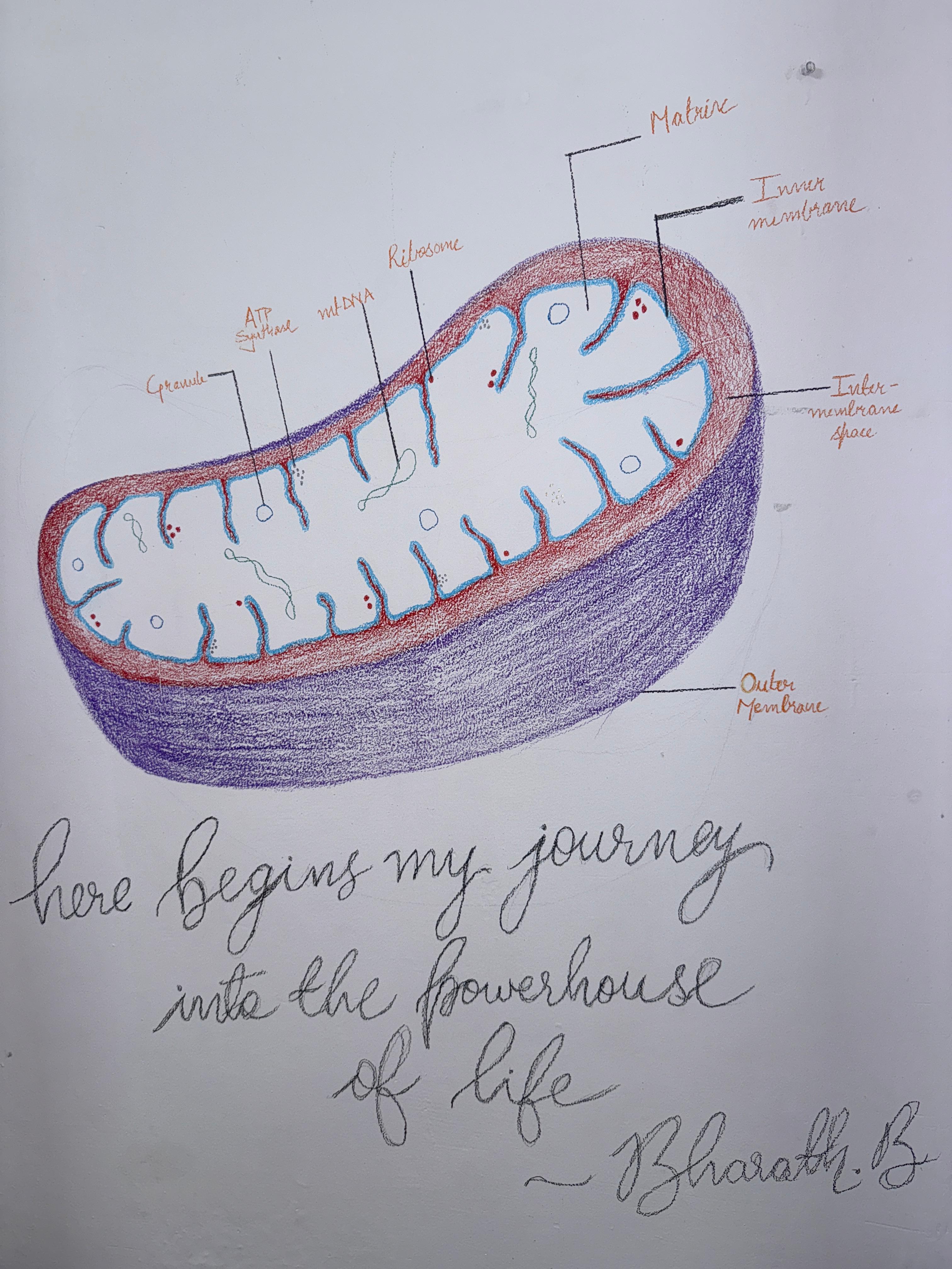
Description: This is not just a drawing, it is a declaration of purpose. I drew this mitochondrion on the wall of my hostel room, where I study, dream, and reflect. Learning about mitochondria was the spark that lit up my curiosity towards science. This drawing represents a commitment for me as I move from textbooks to research, and a reminder of the scientific path I have chosen and of the questions that drive me.
“Why do certain cells lose mitochondria and live briefly (like RBCs), while others retain them and live for years, yet still die when mitochondria fail?"
"How do mitochondria decide the fate of a cell, and how can we intervene before their failure becomes irreversible?"
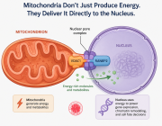
The labelled components of the mitochondrion, ATP synthase, mtDNA, ribosomes, the matrix, and inner membrane, all hold answers we are yet to fully uncover. Art and science both require imagination, structure and a willingness to search for truth.This artwork expresses my passion and dedication to mitochondria, not just as an organelle, but as a central force in life and discovery.
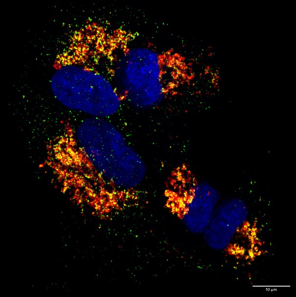
Description : The Super resolution confocal image represents a co-localization between mitochondria (green) and cytochrome c (red) due to Peptidase M84 treatment in PA-1 and SKOV3 ovarian cancer cells. The nucleus was stained with DAPI. Scale Bar 10 μm.
Context of the Study : Mitochondrial cytochrome functions as an electron carrier in the respiratory chain, translocates to the cytosol in Peptidase M84 treated cells undergoing apoptosis, where it participates in the activation of specific caspases. We found an increase in the cytochrome c expression levels in cytosolic fraction devoid of mitochondria in translational levels in Peptidase M84 treated PA-1 and SKOV3 cells. This study leads to the activation of the intrinsic pathway of apoptosis in ovarian cancer cells by Peptidase M84 from B. altitudinis.
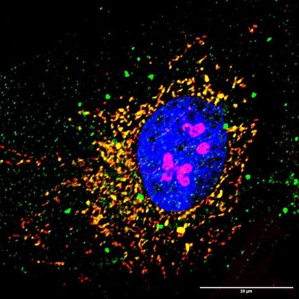
By Subhradip Nath, Senior Researcher from Biophysics and Structural Genomics Division, Saha Institute of Nuclear Physics, Kolkata, India

Description: This Z-projected confocal microscopy image of a C2C12 myoblast reveals the intricate spatial relationship between mitochondria (Red, labeled with MitoTracker™ CMXRos), F-actin (Green, stained with phalloidin), and the nuclear envelope (Blue, marked by lamin A). A striking feature is the dense perinuclear accumulation of mitochondria, suggesting functional compartmentalization around the nucleus. This organization is biologically significant, as perinuclear mitochondria support localized ATP production for nuclear processes, buffer calcium exchange between the ER and mitochondria, and modulate redox signaling that can influence chromatin dynamics and transcription. The actin cytoskeleton appears to coordinate this distribution, underscoring its role in subcellular organelle positioning and metabolic regulation in muscle progenitor cells.
Context of Study: Mitochondrial positioning and function are tightly regulated by the cytoskeleton and nuclear architecture. Lamin A at the nuclear envelope regulates the actin cytoskeleton via the LINC complex, influencing nuclear-cytoplasmic mechanical signalling and cytoskeletal crosstalk. Actin filaments facilitate mitochondrial dynamics, including transport and fission–fusion processes via mitochondrial associated actins or MAA, essential for bioenergetics regulation and energy distribution. In C2C12 myoblasts, the observed perinuclear clustering of mitochondria reflects this coordination, supporting localized ATP production, calcium buffering, and redox signaling in the periphery of the nucleus. Disruption in lamin A alters cytoskeletal integrity and mitochondrial organization, highlighting a critical interplay underlying cellular energy homeostasis and potentially contributing to laminopathies such as dilated cardiomyopathy.
Image 17 - Silent Guardians : Mitochondria in Life and Death
By Krishna Kumar Ganta, Postdoctoral Research Associate at the UMass Chan Medical School, Worcester, USA
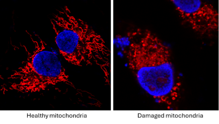
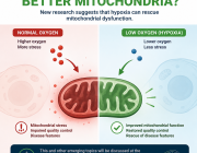
Description of the Image: This image captures two extremes of mitochondrial life: On the left, healthy, interconnected mitochondria form elegant networks, tirelessly fueling the cell's life, metabolism, and health. On the right, fragmented and damaged mitochondria mark the cell's decision to embrace death not violently, but through a well-orchestrated process that limits harm to surrounding cells. Mitochondria sustain us quietly, reflected in our blood work and health, often unnoticed. But when cells reach their limits, mitochondria lead the way toward programmed cell death, preserving the tissue and ensuring minimal collateral damage. This image is a visual tribute to their dual role in life and in death.
Context of the Study: This image originated from experiments assessing mitochondrial damage following antiretroviral therapy (ART) exposure. While focused on drug-induced stress, it reflects a universal biological truth: mitochondria are not just energy providers. They are the gatekeepers of cell fate, enabling life with resilience and guiding death with dignity.
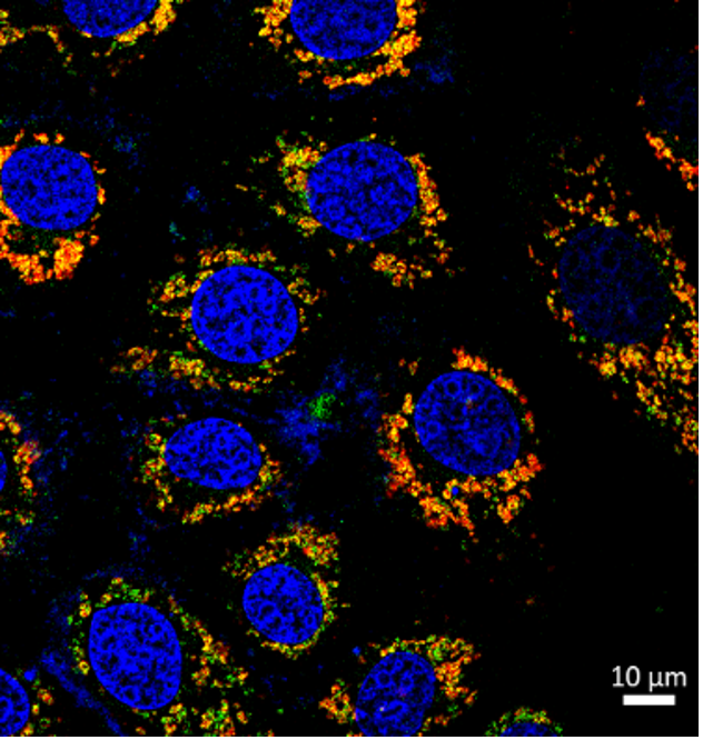
Image 18 - The Fire Behind Every Hearbeat
By Samantha Gallero-San Martin, PhD Student at the ACCDiS; Faculty of Chemical and Pharmaceutical Sciences, University of Chile, Department of Nutrition, Exercise and Sports (NEXS), University of Copenhagen, Denmark.

Description: This live-cell image shows the detailed architecture of an isolated adult mouse cardiomyocyte. Actin filaments (grey) provide structural support, while mitochondria (orange) are distributed throughout the cell, reflecting their critical role in meeting the high energy demands required for cardiac contraction. This visualization highlights the complex spatial interface between the cytoskeleton and mitochondria on maintaining heart beats.
Context: Mitochondria are essential for cardiac function, generating over 90% of the ATP required to fuel continuous contraction and relaxation cycles. In cardiomyocytes, their distribution and interaction with the cytoskeleton support efficient energy transfer and localized signaling critical for maintaining contractile performance. This imaging approach allows us to examine these organelle networks in living cells, providing insights into how metabolic stress, such as caloric overload and insulin resistance, may disrupt mitochondrial organization and function.
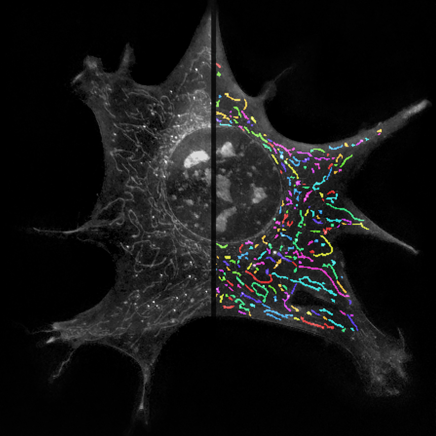
Image 19 - Living Networks: Preserving Mitochondrial Health with Label-free Holotomographic Vision
Submit your Image and win a Free Registration
Have a memorable mitochondria image?
Submit your image for a chance to win free in-person or virtual registration to the 2025 congress.
We also welcome original artwork or drawings related to mitochondria, whether it's about life, energy, or dynamics.
Image Submission Guidelines:
To enter the contest, please submit the following information in a Word document:
- Your name
- Your complete affiliation
- A picture of yourself (optional)
- Your mitochondria image, including: A title/ A description of the image/ The context of the study
How to Enter?
Send your submissions to: mitochondria[at]wms-site.com.
Share your vision of mitochondria and stand out!