
Transplantation of Astrocytic Mitochondria for Intracerebral Hemorrhage Treatment
- Details
- Published on 17 August 2022


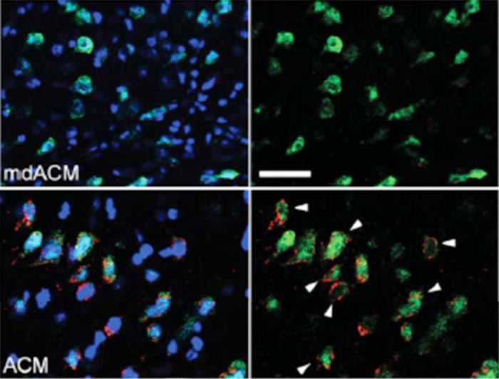
Level of Mn-SOD in neurons 21 days after hemmorage in mice that received mitochondria with and without Mn-SOD at one hour, seven days, and 14 days post-hemmorage. White arrows indicate the cells showing high Mn-SOD (red) in the neurons as a result of the treatment.
News Release, World Mitochondria Society, Berlin - Germany – August 17, 2022
Astrocytes release functional mitochondria that play regulatory and pro-survival functions upon entering adjacent cells. These released mitochondria can enter microglia and promote their reparative/pro-phagocytic phenotype that assists in hematoma cleanup and neurological recovery after intracerebral hemorrhage.
The robust increase in superoxide generation and elevated oxidative damage during intracerebral hemorrhage coincides with loss of the mitochondrial enzyme manganese superoxide dismutase (Mn-SOD). Tahiro et al. showed that intravenous transplantation of astrocytic mitochondria reversed the damaging effect of intracerebral hemorrhage.
When the astrocytic mitochondria entered the brain (and neurons) they restored Mn-SOD levels and reduced neurological deficits in male mice subjected to intracerebral hemorrhage. Astrocytic mitochondria prevented reactive oxygen species-induced oxidative stress and neuronal death by restoring neuronal Mn-SOD levels, while at the same time promoted neurite extension and upregulation of synaptogenesis-related gene expression. The mitochondria genome-encoded small peptide humanin could simulate mitochondria-transfer effect on neuronal Mn-SOD expression, oxidative stress, and neuroplasticity under intracerebral hemorrhage-like injury.
In summary, adoptive astrocytic mitochondrial transfer enhances neuronal Mn-SOD-mediated anti-oxidative defense and neuroplasticity in the brain, which potentiate functional recovery following intracerebral hemorrhage.
© Image -Tashiro et al., JNeurosci 2022
Targeting Mitochondria 2022 will dedicate a whole session for Mitochondrial transplantation and transfer. Conference Program.
Media contact:
World Mitochondria Society
This email address is being protected from spambots. You need JavaScript enabled to view it.
+33-1-5504-7755
Targeting Mitochondria 2022 Congress
October 26-28, 2022 - Berlin, Germany
wms-site.com
Researchers prove the potential of mitochondria-targeted chemotherapies
- Details
- Published on 06 July 2022
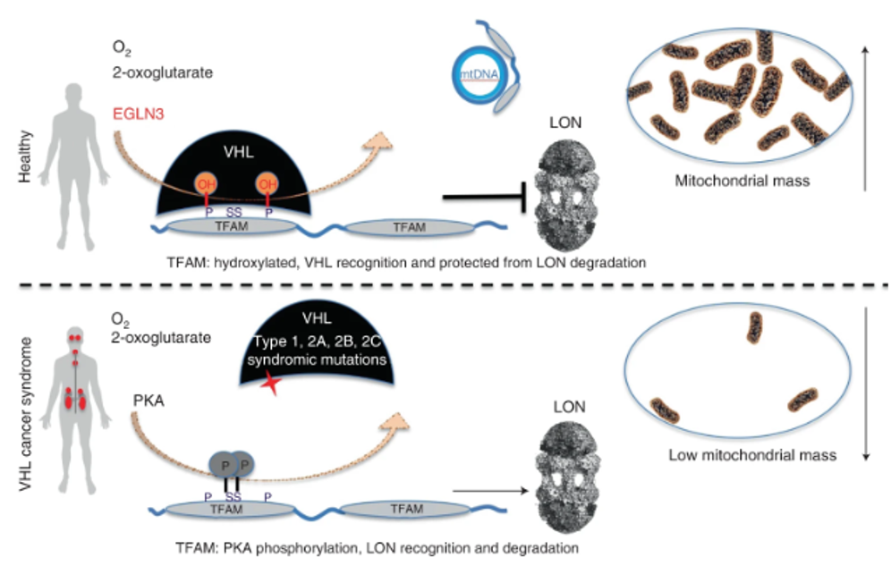
Schematic of oxygen-dependent regulation of mitochondrial transcription factor TFAM by pVHL, independent of the canonical substrate HIFα. mtDNA, mitochondrial DNA
News Release, World Mitochondria Society, Berlin - Germany – July 6, 2022
Researchers at Karolinska Institutet in Sweden have linked resistance to treatment for a deadly form of kidney cancer to low mitochondrial content in the cell. When the researchers increased the mitochondrial content with an inhibitor, the cancer cells responded to the treatment. Their findings, which are published in Nature Metabolism, offer hope for more targeted cancer drugs.
Mitochondria produce energy for the cell and require oxygen to do so. As such, they are the most oxygen-demanding component of the cell. But how mitochondria adapt in a low-oxygen environment and are linked to cancer therapy resistance has remained unknown.
“We’ve shown for the first time how the formation of new mitochondria is regulated in cells that lack oxygen and how this process is altered in cancer cells with VHL mutations,” says Associate Professor Susanne Schlisio, group leader at the Department of Microbiology, Tumor and Cell Biology, Karolinska Institutet.
Healthy cells are prevented from becoming cancerous by a gene called von Hippel-Lindau (VHL). The 2019 Nobel Prize in Physiology or Medicine was awarded to the discovery that VHL was part of the cell’s oxygen detection system. Normally, VHL breaks down another protein called HIF. Consequently, when VHL is mutated, HIF accumulates and causes a disease called VHL syndrome in which the cells react as if they lack oxygen despite oxygen being present. VHL syndrome greatly increases the risk of tumours, both benign and malignant. VHL syndrome-induced kidney cancer has a poor prognosis, with a five-year survival rate of barely 12 per cent.
In the present study, the researchers examined the protein content of cancer cells from patients with different variants of VHL syndrome, and how they differed from another group of individuals with a special VHL mutation called Chuvash, a mutation involved in hypoxia-sensing disorders without any tumor development. Those with the Chuvash VHL-mutation had normal mitochondria in their cells, while those with VHL syndrome mutation had few.
To increase the amount of mitochondrial content in VHL related kidney cancer cells, the researchers treated these tumours with an inhibitor of a mitochondrial protease called “LONP1”. The cells then became susceptible to the cancer drug sorafenib, which they had previously resisted. In mouse studies, this combination treatment led to reduced tumour growth.
“We hope that this new knowledge will pave the way for more specific LONP1 protease inhibitors to treat VHL-related clear cell kidney cancer,” says the study’s first author Shuijie Li, postdoctoral researcher in the Schlisio’s group. “Our finding can be linked to all VHL syndromic cancers, such as the neuroendocrine tumours pheochromocytoma and paraganglioma, and not just kidney cancer.”
Targeting Mitochondria 2022 will cover all recent advances in the implication of mitohcondria in chronic diseases, as well as in the acceleration of mitochondrial medicine. If you are currently working on these topics, you can submit your abstract.
Media contact:
World Mitochondria Society
This email address is being protected from spambots. You need JavaScript enabled to view it.
+33-1-5504-7755
Targeting Mitochondria 2022 Congress
October 26-28, 2022 - Berlin, Germany
wms-site.com
Near-Infrared Light Modulation of Cytochrome C Oxidase in Traumatic Brain Injury
- Details
- Published on 30 June 2022
News Release, World Mitochondria Society, Berlin - Germany – June 30, 2022
Traumatic brain injuries (TBIs) due to sudden impact to the head alter behavior and impair physical and cognitive function. The outcome of TBI is influenced by the patient’s biological sex besides the severity, type and the area of the brain affected.
Literature on mitochondrial dysfunction mainly focused on exponential reactive oxygen species (ROS) generation, increased mitochondrial membrane potential, and altered mitochondrial dynamics as a key player in the outcome to brain injury.
In this study they evaluated the effect of a near-infrared (NIR) light exposure on gene expression in a Drosophila TBI model. NIR interacts with cytochrome c oxidase (COX) of the electron transport chain to reduce mitochondrial membrane potential hyperpolarization, attenuate ROS generation, and apoptosis.
They subjected w1118 male and female flies to TBI using a high-impact trauma (HIT) device and subsequently exposed the isolated fly brains to a COX-inhibitory wavelength of 750 nm for 2 hours. Genome-wide 3′-mRNA-sequencing of fly brains revealed that injured w1118 females exhibit greater changes in transcription compared to males at 1, 2, and 4 hours after TBI.
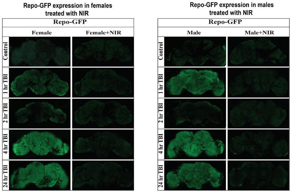
Confocal images showing change in GFP expression in Repo-GFP flies at control, 1, 2, 4, and 24 hr after TBI in females and males
Inhibiting COX by exposure to NIR downregulated gene expression in injured females but had minimal effect in injured males. Those obtained results suggested that mitochondrial COX modulation with NIR alters gene expression in Drosophila following TBI and the response to injury and NIR exposure varies by biological sex.
Targeting Mitochondria 2022 will specialize a session on "Translational Therapies - Focus on Infrared Therapies" chaired by a where a memeber of this research team Dr. Maik Hüttemann.
Remember that the scientific committee will welcome your abstracts related to this hot topic. Submit abstract.
Media contact:
World Mitochondria Society
This email address is being protected from spambots. You need JavaScript enabled to view it.
+33-1-5504-7755
Targeting Mitochondria 2022 Congress
October 26-28, 2022 - Berlin, Germany
wms-site.com
Parkin in the Regulation of Myocardial Mitochondria-Associated Membranes and Cardiomyopathy During Endotoxemia
- Details
- Published on 30 June 2022
News Release, World Mitochondria Society, Berlin - Germany – June 30, 2022
Mitochondrial deficiency is a known pathology in sepsis-induced organ failure. Qun Sophia Zang and the team had previously reported that the mitochondria-associated membranes (MAMs), a subcellular domain supporting mitochondrial status, are impaired in the heart during endotoxemia, suggesting a mechanism of mitochondrial damage occurred in sepsis. Mitophagy pathway via E3 ubiquitin ligase Parkin and PTEN-induced kinase 1 (PINK1) controls mitochondrial quality. Studies described here examined the impact of Parkin on cardiac MAMs and endotoxemia-induced cardiomyopathy. Additionally, point mutation W403A in Parkin was previously identified as a constitutively active mutation in vitro. In vivo effects of forced expression of this mutation were evaluated in the endotoxemia model.
In this study, mice of wild type (WT), Parkin-deficiency (Park2 −/− ), and knock-in expression of Parkin W402A (human Parkin W403A) were given lipopolysaccharide (LPS) challenge. Cardiac function was evaluated by echocardiography. In the harvested heart tissue, MAM fractions were isolated by ultracentrifugation, and their amount and function were quantified. Ultrastructure of MAMs and mitochondria was examined by electron microscopy. Mitochondrial respiratory activities were measured by enzyme assays. Myocardial inflammation was estimated by levels of pro-inflammatory cytokine IL-6. Myocardial mitophagy was assessed by levels of mitophagy factors associated with mitochondria and degrees of mitochondria-lysosome co-localization. Parkin activation, signified by phosphorylation on serine 65 of Parkin, was also evaluated.
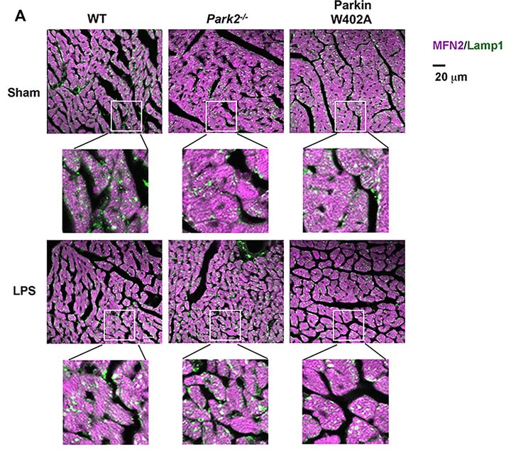
Heart tissue sections were co-stained with antibodies against lysosomal marker Lamp1 (green) and mitochondrial marker Mfn2 (purple). Colors in white and pale green are resulted from co-localization of the two markers.
Compared with WT, Park2 −/− mice showed more severely impaired cardiac MAMs during endotoxemia, characterized by disrupted structure, reduced quantity, and weakened transporting function. Endotoxemia-induced cardiomyopathy was intensified in Park2 −/− mice, shown by worsened cardiac contractility and higher production of IL-6. Mitochondria from the Park2 −/− hearts were more deteriorated, indicated by losses in both structural integrity and respiration function. Unexpectedly, mice carrying Parkin W402A showed similar levels of cardiomyopathy and mitochondrial damage when compared with their WT counterparts. Further, Parkin W402A mutation neither enhanced mitophagy nor increased Parkin activation in myocardium under the challenge of endotoxemia.
As suggested by the results, Parkin/PINK1 mitophagy participates in the regulation of cardiac MAMs during endotoxemia. Point mutation W402A (human W403A) in Parkin is not sufficient to alleviate cardiomyopathy induced by endotoxemia in vivo.
Join us at Targeting Mitochondria 2022 and benefit from the experience of Dr. Qun Sophia Zang in this field. Early Registration.
Media contact:
World Mitochondria Society
This email address is being protected from spambots. You need JavaScript enabled to view it.
+33-1-5504-7755
Targeting Mitochondria 2022 Congress
October 26-28, 2022 - Berlin, Germany
wms-site.com
Molecular Machinery That Delivers Metabolites to Mitochondria
- Details
- Published on 13 May 2022
News Release, World Mitochondria Society, Berlin - Germany – May 13, 2022
Metabolites enter the mitochondria.
The process goes far beyond fueling power generation in the cell, according to Hongying Shen, PhD, assistant professor of cellular & molecular physiology at Yale School of Medicine and a member of Yale’s Systems Biology Institute. “Mitochondria also house many other biochemical processes that are critical for cellular and organismal physiology, and that require trafficking in and out of all kinds of metabolites, including nucleotides, amino acids for protein, and lipids,” she says.
In a study published May 5 in Nature Communications, Shen and her lab have identified the molecular machinery through which many of the metabolites reach inside the mitochondria.
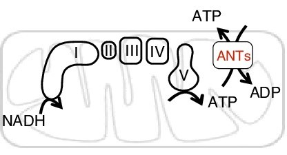
The function of adenine nucleotide carriers in supporting bioenergetics.
They focus on the human SLC25 carrier family, the largest protein family responsible for metabolite translocation across the mitochondrial membrane. Each of the 53 transporters has a distinct assignment. “They are structurally, sequence-wise, very similar to each other,” says Shen, “but they have this amazing specificity. One is dedicated to a certain type of nutrients, the other dedicated to other metabolites or nutrients. So there seems to be a very tight regulation in terms of specificity to recognize metabolites being transported.”
This new knowledge may open the door to potential regulation of what enters the cell, with the goal of preventing or mitigating disease.
A blueprint for treating neurological disorders?
“We are particularly interested in human diseases affecting the brain that include psychiatric disorders and neurodegenerative disorders,” Shen explains. “In fact, there have been de novo mutations in the gene SLC25A39 that have been implicated in autism. And also, A39 has been recently implicated in Parkinson’s disease where oxidative stress was proposed as a pathological mechanism.” In addition, according to Shen, the antioxidant metabolite glutathione, whose delivery route her lab also identified, may be of great interest to scientists studying cancer.
One day in the future, it is conceivable that biomarkers could associate conditions such as neurodegeneration with the metabolic processes that Shen’s lab is studying. That, she says, could lead to new treatments for disease. “Then we can perhaps change our metabolism by diet and by nutrition and all kinds of methods to intervene with that. If we were able to discover these processes and identify the metabolites, can we use dietary intervention to slow the disease onset or disease progression? There's a long way to go [before we might accomplish that], but it's something.”
The new study appears to lay a sound foundation for future work.
Targeting Mitochondria 2022 will cover the most recent studies on role of the mitochondria in health and in disease. It will unravel the latest discoveries and mechanisms in this field with the help of international professional scientists. Meet the speakers.
Media contact:
World Mitochondria Society
This email address is being protected from spambots. You need JavaScript enabled to view it.
+33-1-5504-7755
Targeting Mitochondria 2022 Congress
October 26-28, 2022 - Berlin, Germany
wms-site.com


