
Iron & Cancer : Targeting Mitochondria
- Details
- Published on 08 May 2023


News Release, World Mitochondria Society, Berlin - Germany – May 8, 2023
In their new paper published in Science Advances, Subhadip Mukhopadhyay and colleagues from NYU and Harvard, highlighted a critical link between autophagy, iron metabolism, and mitochondrial function that may have implications for Pancreatic ductal adenocarcinoma (PDAC) progression.
PDAC cells maintain a high level of autophagy, allowing them to thrive in an austere microenvironment. However, the processes through which autophagy promotes PDAC growth and survival are still not fully understood.
Subhadip Mukhopadhyay and his research team showed that autophagy inhibition in PDAC alters mitochondrial function by losing succinate dehydrogenase complex iron sulfur subunit B expression by limiting the availability of the labile iron pool. PDAC uses autophagy to maintain iron homeostasis, while other tumor types assessed require macropinocytosis, with autophagy being dispensable.
The researchers observed that cancer-associated fibroblasts can provide bioavailable iron to PDAC cells, promoting resistance to autophagy ablation. To overcome this cross-talk, they used a low-iron diet and demonstrated that this augmented the response to autophagy inhibition therapy in PDAC-bearing mice.
In summary, this work supports the model in which the iron-autophagy/lysosome axis represents a metabolic vulnerability in PDAC. It specifically demonstrated the feasibility of targeting this metabolic dependency in vivo and mechanistically defined the distinct regulation of iron homeostasis in different tumor types, which has implications for how one would approach this therapeutically.
Targeting Mitochondria 2023 will extensively cover the implication of mitochondria in cancer and its potential in cancer therapy. Submit a related abstract.
Media contact:
World Mitochondria Society
This email address is being protected from spambots. You need JavaScript enabled to view it.
+33-1-5504-7755
Targeting Mitochondria 2023 Congress
October 11-13, 2023 - Berlin, Germany
wms-site.com
Rescuing Corneal Cells from Death with the Help of Mitochondria
- Details
- Published on 20 April 2023

News Release, World Mitochondria Society, Berlin - Germany – April 20, 2023
Fuchs' endothelial corneal dystrophy, a degenerative eye disease, causes progressive vision loss that can induce blindness. It is the leading cause of corneal transplantation, but the scarcity of grafts hinders its treatment. A research team from Université Laval and Université de Montréal has identified a way to slow the disease and even avoid transplantation if diagnosed at an early stage.
In people with the disease, the endothelial cells at the back of the cornea die more quickly than in healthy people.
"Everyone loses them at a slow rate, slow enough to make it to the end of our lives without problems. For sick people, depletion is accelerated by factors not yet understood at the molecular level. Since the cells do not divide, they do not replace themselves," says Patrick J. Rochette, full professor at the Faculty of Medicine at Université Laval and a researcher at the CHU de Québec-Université Laval Research Center, who conducted the study.
These endothelial cells play an essential role in vision. They ensure that the cornea remains transparent by keeping it partially dehydrated. When the cells die, the cornea becomes wet and cloudy, which can lead to complete blindness.
In a previous study, the research team showed that mitochondria were central to the disease. "In people with the disease, the mitochondria become depleted rapidly, leading to cell death. The more cells die, the more the mitochondria in other cells have to compensate, which accelerates their depletion. It's a vicious circle," explains Patrick J. Rochette.
Reduce mortality rate
The research team wondered whether injecting healthy mitochondria into cells could delay the progression of Fuchs' dystrophy. To test their hypothesis, the scientists used diseased endothelium removed during a corneal transplant. "We were able to save cells close to death, going from a 60% mortality rate to 10%," Rochette said. These results demonstrate a high therapeutic potential for the injection of mitochondria.
The strength of this approach lies in the automatic recycling of diseased mitochondria without injecting healthy ones directly into the cell. "Cells eat mitochondria as if their life depended on them. Any cell, even if it is dying, will take them up. The replacement happens by itself. After 24 hours, only healthy mitochondria remain," says the researcher.
In the study, the healthy mitochondria were grown under controlled conditions, but the research group is developing an approach to extract them from the patient's blood.
If Fuchs' dystrophy is diagnosed at an early stage, when most endothelial cells are still alive, the approach could maintain vision without a transplant. The injection of mitochondria would be a benign procedure, much less invasive than surgery.
The paper is published in the journal Scientific Reports.
Targeting Mitochondria 2023 will adress the latest research that targets the mitochondria in ophthalmology. You can submit a related abstract.
Source: Press release by the University of Laval.
Image Credits: Freepik
Media contact:
World Mitochondria Society
This email address is being protected from spambots. You need JavaScript enabled to view it.
+33-1-5504-7755
Targeting Mitochondria 2023 Congress
October 11-13, 2023 - Berlin, Germany
wms-site.com
Key Information About The Function of Mitochondria in Cancer Cells
- Details
- Published on 16 March 2023
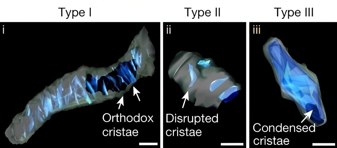
SBEM images and 3D reconstruction of representative type I (i), II (ii) and III (iii) crista structures identified in OXPHOSHI LUAD cells and OXPHOSLO LUSC cells.
News Release, World Mitochondria Society, Berlin - Germany – March 16, 2023
Scientists have long known that mitochondria play a crucial role in the metabolism and energy production of cancer cells. However, until now, little was known about the relationship between the structural organization of mitochondrial networks and their functional bioenergetic activity at the level of whole tumors.
In a new study, published in Nature, researchers from the UCLA Jonsson Comprehensive Cancer Center used positron emission tomography (PET) in combination with electron microscopy to generate 3-dimensional ultra-resolution maps of mitochondrial networks in lung tumors of genetically engineered mice.
They categorized the tumors based on mitochondrial activity and other factors using an artificial intelligence technique called deep learning, quantifying the mitochondrial architecture across hundreds of cells and thousands of mitochondria throughout the tumor.
The authors examined two main subtypes of non-small cell lung cancer (NSCLC)—adenocarcinomas and squamous-cell carcinomas and found distinct subpopulations of mitochondrial networks within these tumors. Importantly, they discovered that the mitochondria frequently organize themselves with organelles such as lipid droplets to create unique subcellular structures that support tumor cell metabolism and mitochondrial activity.
The study was led by Mingqi Han, Ph.D., a post-doctoral researcher in the lab of David Shackelford, Ph.D. Dr. Shackelford is a UCLA Jonsson Comprehensive Cancer Center member and Associate Professor of Pulmonary and Critical Care Medicine at the UCLA David Geffen School of Medicine.
The authors anticipate that mitochondrial populations in human cancer samples will not be mutually exclusive to their respective tumor subtype, but rather there will be a spectrum of activity.
The investigators say these findings provide key information about the function of mitochondria in cancer cells and could lead to new approaches to cancer treatment.
"Our study represents a first step towards generating highly detailed 3-dimensional maps of lung tumors using genetically engineered mouse models," said Dr. Shackelford.
"Using these maps, we have begun to create a structural and functional atlas of lung tumors, which has provided us valuable insight into how tumor cells structurally organize their cellular architecture in response to the high metabolic demands of tumor growth. Our findings hold promise to inform and improve current treatment strategies while illuminating new directions from which to target lung cancer."
"Our study has uncovered a novel finding in the metabolic flux of lung tumors, revealing that their nutrient preference may be determined by the compartmentalization of their mitochondria with other organelles, either relying on glucose ('sugar') or free fatty acids ('fat')," said Dr. Han.
"This discovery has important implications for developing effective anti-cancer therapies that target tumor-specific nutrient preferences. Our multi-modality imaging approach has enabled us to uncover this previously unknown aspect of cancer metabolism, and we believe that it can be applied to other types of cancer, paving the way for further research in this area."
Image Credit: David Shackelford et al, Nature (2023)
Targeting Mitochondria 2023 Conference will elaborate on the latest mitochondria and cancer research and discoveries. You can submit your related abstracts here.
Media contact:
World Mitochondria Society
This email address is being protected from spambots. You need JavaScript enabled to view it.
+33-1-5504-7755
Targeting Mitochondria 2023 Congress
October 11-13, 2023 - Berlin, Germany
wms-site.com
Novel drug makes mice skinny even on sugary, fatty diet
- Details
- Published on 03 April 2023
News Release, World Mitochondria Society, Berlin - Germany – April 3, 2023
Researchers from The University of Texas Health Science Center at San Antonio (UT Health San Antonio) have developed a small-molecule drug that prevents weight gain and adverse liver changes in mice fed a high-sugar, high-fat Western diet throughout life.
"When we give this drug to the mice for a short time, they start losing weight. They all become slim," said Madesh Muniswamy, PhD, professor of medicine in the health science center's Joe R. and Teresa Lozano Long School of Medicine.
Findings by the collaborators, also from the University of Pennsylvania and Cornell University, were published Feb. 27 in the high-impact journal Cell Reports. Muniswamy, director of the Center for Mitochondrial Medicine at UT Health San Antonio, is the senior author.
Fourth most common element
The research team discovered the drug by first exploring how magnesium impacts metabolism, which is the production and consumption of energy in cells. This energy, called ATP, fuels the body's processes.
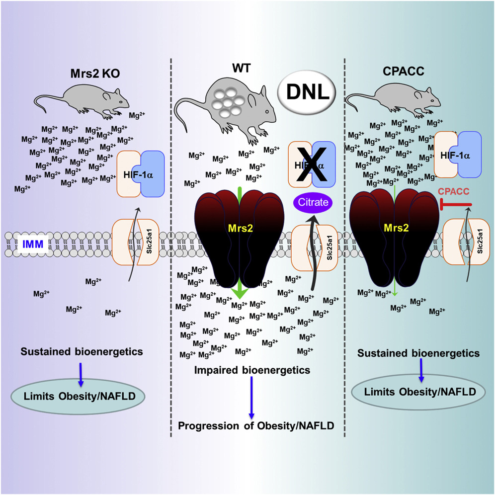
Magnesium is the fourth most abundant element in the body after calcium, potassium and sodium, and plays many key roles in good health, including regulating blood sugar and blood pressure and building bones. But the researchers found that too much magnesium slows energy production in mitochondria, which are cells' power plants.
"It puts the brake on, it just slows down," said co-lead author Travis R. Madaris, doctoral student in the Muniswamy laboratory at UT Health San Antonio.
Deleting MRS2, a gene that promotes magnesium transport into the mitochondria, resulted in more efficient metabolism of sugar and fat in the power plants. The result: skinny, healthy mice.
Liver and adipose (fat) tissues in the rodents showed no evidence of fatty liver disease, a complication related to poor diet, obesity and type 2 diabetes.
Small-molecule agent
The drug, which the researchers call CPACC, accomplishes the same thing. It restricts the amount of magnesium transfer into the power plants. In experiments, the result was again: skinny, healthy mice. UT Health San Antonio has filed a patent application on the drug.
The mice served as a model system of long-term dietary stress precipitated by the calorie-rich, sugary and fatty Western diet. The familiar results of this stress are obesity, type 2 diabetes and cardiovascular complications.
"Lowering the mitochondrial magnesium mitigated the adverse effects of prolonged dietary stress," said co-lead author Manigandan Venkatesan, PhD, postdoctoral fellow in the Muniswamy lab.
Joseph A. Baur, PhD, of the University of Pennsylvania and Justin J. Wilson, PhD, of Cornell are among the collaborators. "We came up with the small molecule and Justin synthesized it," Madaris said.
Significant implications
"These findings are the result of several years of work," Muniswamy said. "A drug that can reduce the risk of cardiometabolic diseases such as heart attack and stroke, and also reduce the incidence of liver cancer, which can follow fatty liver disease, will make a huge impact. We will continue its development."
Funders of this project include the National Institutes of Health, the U.S. Department of Defense and the San Antonio Partnership for Precision Therapeutics.
Image Credit: Madaris, Travis R. et al. Cell Reports (2023)
Targeting Mitochondria 2023 Conference will elaborate on the latest mitochondria targeting drugs. You can submit your related abstracts here.
Media contact:
World Mitochondria Society
This email address is being protected from spambots. You need JavaScript enabled to view it.
+33-1-5504-7755
Targeting Mitochondria 2023 Congress
October 11-13, 2023 - Berlin, Germany
wms-site.com
Researchers discover therapeutic target to aid in glaucoma treatment
- Details
- Published on 13 March 2023
News Release, World Mitochondria Society, Berlin - Germany – March 13, 2023
Indiana University School of Medicine researchers have identified a new therapeutic target that could lead to more effective treatment of glaucoma.
Glaucoma is a neurodegenerative disease that causes vision loss and blindness due to a damaged optic nerve. More than 200,000 people are affected by glaucoma in the United States each year. Unfortunately, there is currently no treatment. In a newly published paper in Communications Biology, researchers found neurons use mitochondria for a steady source of energy, and restoring mitochondrial homeostasis in the diseased neurons can protect the optic nerve cells from being damaged.
“Age-related neurodegenerative disease, which includes glaucoma, Parkinson’s disease, and amyotrophic lateral sclerosis (ALS), is the biggest global health problem,” said Arupratan Das, PhD, assistant professor of ophthalmology and principal investigator of the study. “The fundamental mechanisms that we discovered can be used to protect neurons in glaucoma and be tested for the other diseases. We have identified a critical step of complex mitochondrial homeostasis process, which rejuvenates the dying neuron, similar to giving a lifeline to a dying person.”
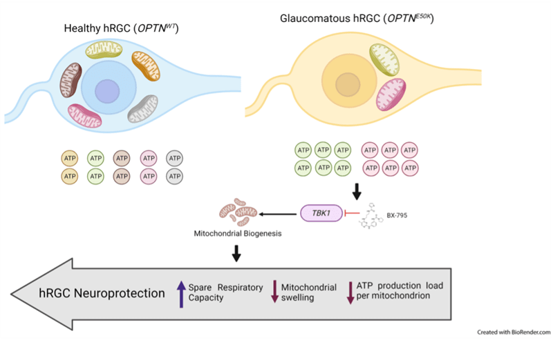
The research team, led by Michelle Surma and Kavitha Anbarasu from the Department of Ophthalmology, used induced pluripotent stem cells (iPSCs) from patients with and without glaucoma as well as clustered regularly interspaced short palindromic repeats (CRISPR) engineered human embryonic stem cells with glaucoma mutation. Using stem cell differentiated retinal ganglion cells (hRGCs) of the optic nerve, electron microscopy and metabolic analysis, researchers identified glaucomatous retinal ganglion cells suffer mitochondrial deficiency with more metabolic burden on each mitochondrion. This leads to mitochondrial damage and degeneration. Mitochondria are the tube like structures in cells which produce adenosine triphosphate, cell’s energy source.
However, the process could be reversed by enhancing mitochondrial biogenesis by a pharmacological agent. The team showed retinal ganglion cells are highly efficient in degrading bad mitochondria, but at the same time producing more to maintain homeostasis.
“Finding that retinal ganglion cells with glaucoma produce more adenosine triphosphate even with less mitochondria was astonishing,” Das said. “However, when triggered to produce more mitochondria, the adenosine triphosphate production load was distributed among more mitochondrion which restored the organelle physiology. It is similar to a situation where a heavy stone is carried by fewer people versus a greater number of people—each person will have less pain and injury, just like each mitochondrion will have less difficulty and damage.”
In the future, Das would like to test if these mechanisms protect the optic nerve in animal models under injury before testing in humans to hopefully lead to new clinical interventions.
Targeting Mitochondria 2023 Conference will elaborate on the latest mitochondria research and discoveries. You can submit your related abstracts here.
Media contact:
World Mitochondria Society
This email address is being protected from spambots. You need JavaScript enabled to view it.
+33-1-5504-7755
Targeting Mitochondria 2023 Congress
October 11-13, 2023 - Berlin, Germany
wms-site.com


