
Enhancing T Cell Antitumor Activity through Mitochondrial Transfer
- Details
- Published on 18 September 2024


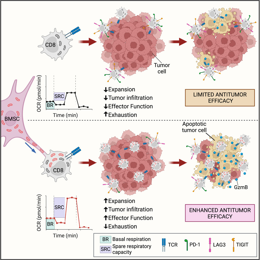
The research demonstrated that bone marrow stromal cells (BMSCs) transfer mitochondria to T cells through intercellular nanotube connections, significantly increasing T cell mitochondrial mass and respiration. This process, requiring Talin 2 on both donor and recipient cells, improves the metabolic function of T cells, enabling them to infiltrate tumors more effectively and show fewer signs of exhaustion.
By utilizing this intercellular mitochondrial transfer, the study offers a new approach to overcoming T cell exhaustion, a critical challenge in immunotherapy. This breakthrough in organelle medicine could lead to the development of next-generation cell therapies with enhanced efficacy against cancer.
The latest innovations in mitochondrial research and mitochondrial transfer, including findings like this, will be discussed at the 15th World Congress on Targeting Mitochondria, taking place from October 29-31, 2024, in Berlin, Germany.Image Credits: Graphical Abstract Baldwin, Jeremy G. et al. Cell (2024)
Mitochondria: Key Players in Autism Spectrum Disorder?
- Details
- Published on 06 September 2024
Recent review from the Hebrew University of Jerusalem highlighted the multifaceted role of mitochondria in Autism Spectrum Disorder (ASD), suggesting that mitochondrial dysfunction may significantly contribute to the development and pathology of this neurodevelopmental disorder. Mitochondria, essential for producing the aerobic energy necessary for brain function, have been found to exhibit abnormalities in individuals with ASD, which could profoundly impact brain development and function.
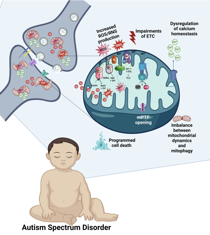
The brain displays high mitochondrial content, particularly in the synapses (shown in the upper left part of the figure). Increased mitochondrial levels of ROS, NO, and RNS, ETC impairments leading to the breakdown of OXPHOS and ATP production, dysregulation of the mitochondrial Ca2+cycling, imbalance between mitochondrial dynamics and mitophagy, prolonged opening of the mPTP, and activation of various mitochondria-related programmed cell death pathways, all contribute to the synaptic dysfunction and ASD. OXPHOS complexes are shown on the upper semisphere of the mitochondrion: I, NADH dehydrogenase; II, succinate dehydrogenase; III, ubiquinone cytochrome c oxidoreductase; IV, cytochrome c, cytochrome oxidase; and V, ATP synthase. Complexes I-IV belong to ETC.
ASD is associated with a variety of mitochondrial abnormalities, including impaired respiratory function, disrupted calcium (Ca2+) cycling, altered production of reactive oxygen and nitrogen species (ROS/RNS), and issues with the opening of the mitochondrial permeability transition pore (mPTP). These dysfunctions can lead to the activation of various mechanisms of programmed cell death, an imbalance in mitochondrial fusion, fission, and autophagy processes, and disturbances in synaptogenesis and synaptic transmission,all of which affect brain development and may result in behavioral deficits.
The importance of mitochondria in ASD cannot be overstated. These organelles are crucial for numerous cellular functions and can be affected by different pathogenic factors, which may explain the similarity in behavioral phenotypes seen in ASD cases of varying origins. Synapses, along with mitochondria, are considered key players in the molecular mechanisms related to ASD. The convergence of various neurodevelopmental pathological processes on synapses may partly explain the behavioral similarities observed in individuals across the autism spectrum.
Interestingly, as the recent review discusses, synaptic abnormalities are closely tied to mitochondrial dysfunction in ASD, suggesting that mitochondria-associated synaptic disturbances could present robust therapeutic targets that have yet to be explored. Although there are still many unknowns in the mitochondria-related mechanisms of autism, understanding these “blank spots” could pave the way for novel and effective treatments for ASD. This is particularly significant given the increasing prevalence of ASD and the current lack of effective pharmacological treatments.
Figure credits: Khaliulin, I., Hamoudi, W. & Amal, H. Mol Psychiatry (2024).
Advances in Mitochondrial Modulation: How Infrared Light is Changing Brain Injury Recovery
- Details
- Published on 22 August 2024
A recent study has spotlighted the transformative role of near-infrared (NIR) light in improving mitochondrial dynamics and quality control, offering new hope for brain injury recovery following cardiac arrest. Dr. Maik Hüttemann, Wayne State University (USA) and active member of the WMS Scientific Board will join Targeting Mitochondria 2024 Congress in Berlin, where he will delve deeper into these findings and discuss the advances in infrared light treatment.
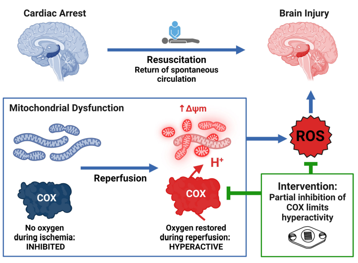
Brain injury remains a significant challenge following cardiac arrest, with mitochondrial dysfunction playing a pivotal role in exacerbating neurological damage. The study investigates how targeting mitochondrial dysfunction with near-infrared light (NIR) wavelengths can mitigate brain injury following cardiac arrest. By employing various models, including isolated porcine brain cytochrome c oxidase (COX), primary mouse neurons, and large animal models, the research provides new insights into NIR-induced mitochondrial modulation.
The research demonstrates that NIR treatment reduces COX activity in an intensity-dependent manner, achieving a controlled modulation of mitochondrial function. This approach results in a moderate reduction of enzyme activity without complete inhibition. Additionally, in neuronal cells, NIR therapy has been shown to decrease mitochondrial swelling and enhance mitophagy, indicating improved mitochondrial health and quality control.
Practical application of NIR therapy has also been investigated. In anesthetized pigs, NIR was found to penetrate deep into the brain with minimal tissue heating, making it a feasible noninvasive treatment option. Moreover, in a model of out-of-hospital cardiac arrest, NIR treatment applied during resuscitation resulted in significantly improved neurological outcomes and reduced brain injury.
The study concludes that NIR effectively modulates mitochondrial function, enhancing mitochondrial dynamics and quality control after ischemia/reperfusion. This noninvasive technique offers promising potential for improving neurological recovery in patients resuscitated from cardiac arrest.
Join Dr. Hüttemann at the Targeting Mitochondria 2024 Congress in Berlin to know more about these findings and explore the future of mitochondria and photomedicine.
Image credits: Wider, J.M., Gruley, E., Morse, P.T. et al. Modulation of mitochondrial function with near-infrared light reduces brain injury in a translational model of cardiac arrest. Crit Care27, 491 (2023).
Mitochondrial DNA in Our Brains Could Be Shortening Lifespan, New Study Reveals
- Details
- Published on 27 August 2024
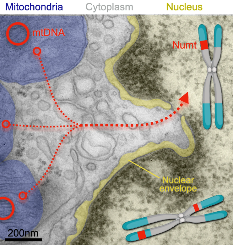
Mitochondria release segments of mitochondrial DNA that can travel through pores of the nucleus and integrate into a cell’s chromosomes (where the insertions are called NUMTs, for nuclear mitochochondrial segments). A new study has found that nuclear mitochondrial DNA insertion—once thought rare—happens in the human brain likely several times over during a person’s lifespan. Credit: Martin Picard laboratory at Columbia University Vagelos College of Physicians and Surgeons
New Treatment Shows Promise for POLG-Related Mitochondrial Disorders
- Details
- Published on 31 July 2024
A clinical trial led by the Research Institute of the McGill University Health Centre (RI-MUHC) has identified Deoxycytidine/Deoxythymidine Combination Therapy as a safe and potentially effective treatment for POLG-related mitochondrial disorders. These disorders cause severe neurological decline, with patients typically surviving only five months after symptom onset. The preliminary results, published in eClinicalMedicine, were largely funded by the Liam Foundation, established after a patient’s diagnosis at the Montreal Children’s Hospital (MCH).
Dr. Kenneth Myers, a pediatric neurologist at MCH, noted, “Our study offers new hope, transforming what was once a death sentence into a chance for a better life. While not a cure, the treatment has significantly improved patients' conditions”.
Understanding the Condition
Mitochondrial diseases, affecting one in 5,000 people, result from dysfunctional mitochondria, the energy-producing parts of cells. In POLG-related disorders, mutations in the POLG gene reduce mitochondrial DNA (mtDNA), leading to seizures, vision loss, muscle issues, nerve damage, developmental delays, and liver failure. The therapy aims to replenish the mtDNA, enhancing mitochondrial function.
After six months, patients showed improved scores on the Newcastle Mitochondrial Disease Scale and lower levels of GDF-15, a marker of mitochondrial dysfunction. Caregivers reported better energy, motor skills, cognition, and communication. No serious side effects were observed.
Dr. Myers highlighted, “Many patients regress dramatically after infections or other triggers. This treatment supplies the mitochondrial DNA they need to function normally.”
Expanding the Trial
The trial included 10 children and adolescents with POLG mutations from the US, Brazil, and India. They received the treatment for six months, with some continuing for 24 months due to significant improvements. Another 14 patients have joined, and a follow-up study on long-term effects is underway.
Liam’s Story: From Despair to Hope
Liam, a ten-year-old with POLG-related mitochondrial disease, began having seizures in 2019. His father, Kevin Reason, started the Liam Foundation after learning about the potential of Deoxycytidine/Deoxythymidine. “Liam is now walking, communicating, and smiling. This treatment gives us hope and vital time to find a cure”, Kevin said.
Liam was the first North American patient in the trial. Thanks to the Liam Foundation and other supporters, 23 more POLG patients have since enrolled.
More Articles...
- Prof. Volkmar Weissig's Interview: Is it time for mitochondria to take centre stage?
- Bridging In Vitro and In Vivo for Mitochondrial Transplantation in Acute Diseases
- Mitochondrial DNA: A Key Player in Cell Death and Inflammation
- Mitochondrial Extracellular Vesicles: Promising Therapeutic Strategy for Neurological Diseases


