
Zika Virus NS1 Hijacks Mitochondria Through Tunneling Nanotubes for Immune Evasion
- Details
- Published on 16 April 2025


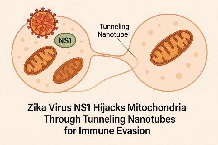
The study titled “Zika virus NS1 drives tunneling nanotube formation for mitochondrial transfer and stealth transmission in trophoblasts” (Nature Communications, February 2025), published by Dr. Indira Mysorekar and her team, explores how Zika virus (ZIKV) utilizes a stealth mechanism to spread among placental trophoblasts. The research focuses on the role of the viral protein NS1 in facilitating this process.
Key Findings:
- Tunneling Nanotube Formation: ZIKV induces the formation of tunneling nanotubes (TNTs) in placental trophoblasts, which serve as conduits for transferring viral components and mitochondria to neighboring uninfected cells.
- Role of NS1 Protein: The non-structural protein 1 (NS1) of ZIKV, particularly its N-terminal 1–50 amino acids, is critical for triggering TNT formation in host cells.
- Immune Evasion: Trophoblasts infected with a TNT-deficient ZIKV mutant elicited a stronger antiviral interferon response compared to those infected with wild-type ZIKV, suggesting that TNT-mediated transmission helps the virus evade host immune defenses.
- Mitochondrial Hijacking: ZIKV infection or NS1 expression leads to increased mitochondrial levels in trophoblasts, with mitochondria being transferred via TNTs from healthy to infected cells, potentially supporting viral replication and survival.
These findings shed light on a novel mechanism by which ZIKV spreads and evades the immune system, offering potential targets for therapeutic intervention.
How Mitochondria Organize Their Powerhouse Machinery for Optimal Performance
- Details
- Published on 09 April 2025

The study titled “In-cell architecture of the mitochondrial respiratory chain,” published in Science on March 20, 2025, presents significant advancements in understanding mitochondrial structure and function.
Key Highlights:
- In Situ Visualization: Researchers employed advanced imaging techniques to observe the mitochondrial respiratory chain within intact cells, providing a detailed view of its native architecture.
- Respiratory Supercomplexes: The study offers insights into how respiratory complexes assemble into supercomplexes, which are crucial for efficient electron transport and energy production in cells.
- Functional Implications: Understanding the organization of these supercomplexes sheds light on their role in cellular metabolism and energy conversion, potentially informing research into mitochondrial-related diseases.
Perspective:
- Challenging previous assumptions: The findings challenge long-standing models that assumed a more fluid, random distribution of respiratory chain components in mitochondrial membranes.
- Biological relevance: By analyzing structures in situ, this study underscores the importance of studying macromolecular organization in native cellular contexts, rather than relying only on purified proteins.
- Broader implications: These insights are critical not only for basic mitochondrial biology but also for understanding mitochondrial dysfunction in aging, neurodegenerative diseases, and metabolic disorders.
- New model for mitochondrial function: This study supports a model in which the geometrical and biochemical compartmentalization within cristae contributes significantly to the efficiency of oxidative phosphorylation.
These findings enhance our comprehension of mitochondrial function and may have implications for addressing metabolic disorders linked to mitochondrial dysfunction.
Why Our Cells Slow Down with Age: The Mitochondria Mystery
- Details
- Published on 02 April 2025
As we get older, our bodies slow down—but have you ever wondered why? New research points to tiny structures inside our cells called mitochondria. These are often called the “powerhouses” of the cell because they produce the energy we need to live. But with age, these powerhouses stop working as well as they used to.
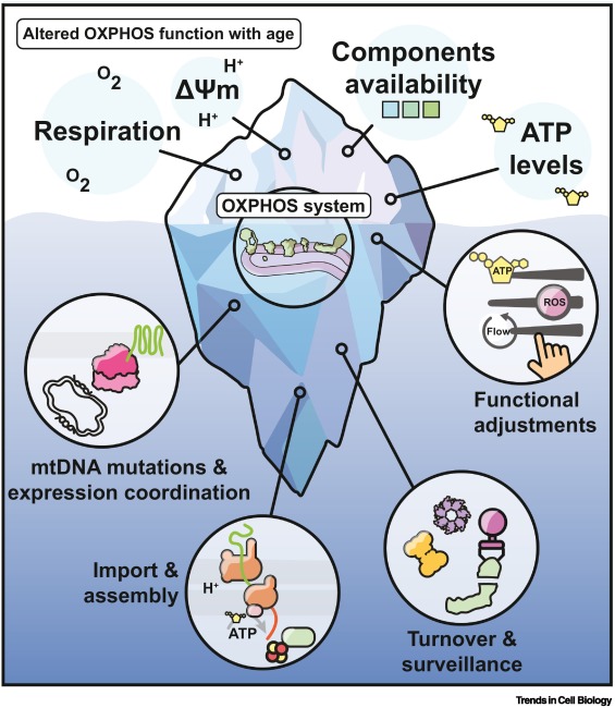
Recent publication by Hanna Salmonowicz et al, have discovered that a key part of the energy-making system, known as OXPHOS, starts to break down over time. This affects how much energy our cells can produce, especially in important organs like the brain, heart, and muscles. It may also explain why we become more vulnerable to diseases as we age.
Main Points:
- Mitochondria need instructions from two sources - our cell’s nucleus and their own DNA—to work properly. With age, this teamwork gets disrupted.
- Mistakes build up in mitochondrial DNA, leading to damage in the energy system.
- Our cells struggle to bring in and assemble the proteins needed to keep mitochondria running.
- Damaged mitochondria make more harmful molecules called ROS (reactive oxygen species), which can hurt the cell even more.
- The effects of these changes are especially strong in parts of the body that use a lot of energy, like the brain and heart.
Conclusion:
This research helps explain why we feel more tired, weaker, and more prone to disease as we age. But it also brings hope. With new tools and technology, scientists are finding ways to better understand and possibly fix these tiny energy factories. Keeping our mitochondria healthy could be the key to living longer, stronger, and healthier lives.
Revolutionary Breakthrough in Mitochondrial Production Offers Hope for Degenerative Diseases
- Details
- Published on 03 April 2025
Scientists have achieved a groundbreaking advancement in the mass production of high-quality human mitochondria, paving the way for transformative treatments in degenerative diseases. By refining stem cell culture conditions, researchers have successfully increased mitochondrial production by an astonishing 854-fold while significantly enhancing their energy output.
Key Highlights:
- Innovative Method: The new method leverages human mesenchymal stem cells and a specially designed culture medium called "mito-condition." This medium integrates nine essential components, including growth factors and human platelet lysate, to optimize mitochondrial production.
- Enhanced Performance: The engineered mitochondria exhibit remarkable therapeutic benefits, notably accelerating cartilage regeneration in osteoarthritis models. They produce 5.7 times more ATP than naturally occurring mitochondria and maintain stable performance even post-isolation.
- Clinical Implications: This breakthrough addresses the long-standing bottleneck in mitochondrial transplantation, which has been constrained by limited supply and inconsistent quality. The ability to produce high-quality mitochondria on a large scale opens new avenues for treating a wide spectrum of mitochondrial dysfunction-related diseases, from joint degeneration to cardiovascular disorders.
- Mechanistic Insights: The study reveals how cells can be reprogrammed to prioritize mitochondrial synthesis by activating the AMPK pathway, a crucial cellular energy sensor. This process downregulates energy-intensive activities like autophagy and secretion, effectively boosting mitochondrial biogenesis.
- Future Applications: Beyond osteoarthritis, this technology holds promise for conditions such as heart disease, neurodegenerative disorders, and wound healing. The concept of organelle tuning could also be adapted to generate other cellular components, broadening the horizons of cell engineering and therapeutic applications.
Publication Details: DOI: 10.1038/s41413-025-00411-6 | Image @WMS
During Targeting Mitochondria 2025, this hot topic will be presented and discussed strongly.
Mitochondria, p53, and the Secret to Aging Gracefully.
- Details
- Published on 24 March 2025
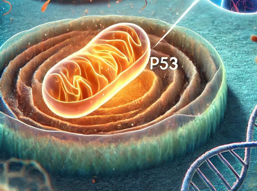
As we age, our cells go through stress and damage—especially to their DNA. One of the key proteins that protects us from this damage is p53, often called the “guardian of the genome.” It helps repair broken DNA and prevents damaged cells from turning into cancer.
But this new study, published in Nature Communications, shows that p53 does even more than we thought: it also helps stop inflammation that can build up in aging cells.
The researchers discovered that mitochondria, which are best known for producing energy, play a key role in controlling how p53 works. When mitochondria function well, they support p53 in repairing DNA and stopping the cell from releasing pieces of damaged DNA—called cytoplasmic chromatin fragments (CCFs)—into the rest of the cell. These fragments can trigger chronic inflammation, a common problem in aging and age-related diseases. By keeping DNA inside the nucleus and helping it stay intact, p53 helps reduce this inflammation and keep the cell stable. When mitochondria aren’t working properly, though, p53 can’t do its job as well—leading to more DNA damage, more CCFs, and more inflammation.
This discovery adds an important piece to the puzzle of aging: it shows how mitochondria and p53 work together to protect cells, maintain DNA integrity, and prevent harmful inflammation. It could open new doors for therapies targeting aging, inflammation, and mitochondrial diseases.
More scientific Details :
Using mitochondrial stress models and p53 loss-of-function systems, the authors show that functional p53:
• Promotes DNA damage repair,
• Limits DNA leakage into the cytoplasm,
• Suppresses cGAS-STING-mediated inflammatory signaling,
• And overall contributes to nuclear genome stability in senescent cells.
Notably, the depletion of p53 led to an increase in nuclear envelope instability and accumulation of CCFs, while its presence correlated with reduced senescence-associated secretory phenotype (SASP) expression. These effects were tightly linked to mitochondrial status, suggesting a mitochondria-to-nucleus signaling axis regulating p53 activity.
This work positions p53 as a key integrator of mitochondrial signals to modulate nuclear architecture and inflammatory outcomes during senescence. It highlights potential therapeutic avenues to modulate mitochondrial-p53 interactions in aging-related pathologies and chronic inflammatory conditions.


