Nominations for The Best Mitochondria Image 2022


- Details
- Published on 11 December 2022
The World Mitochondria Society Scientific Committee will keep accepting images until the end of December 2022! Submit a memorable Mitochondria Image you’ve taken this past year and get the chance to win a free registration for Targeting Mitochondria 2023. Image submissions guidelines.
Images will be shared on the World Mitochondria Society Linked In page. You can vote for your favorite image by pressing the like button. You can find all the nominated images below.
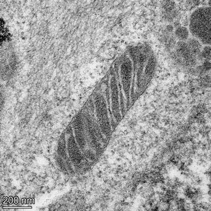
Mitochondrial Crystals
By Catherine Griffiths, Biomedical Imaging Unit Southampton
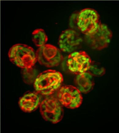
Mitochondrial fireworks – The Ring of Fire
By Simon Licht-Mayer, Institute of Molecular Biotechnology of the Austrian Academy of Sciences
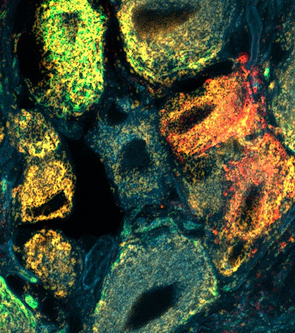
Dorsal root ganglia neurons stained for Neurofilament heavy chain (NF200, green), isolectin B4 (IB4, red), calcitonin gene-related peptide (CGRP, cyan), cytochrome c oxidase I (COX-I, orange)
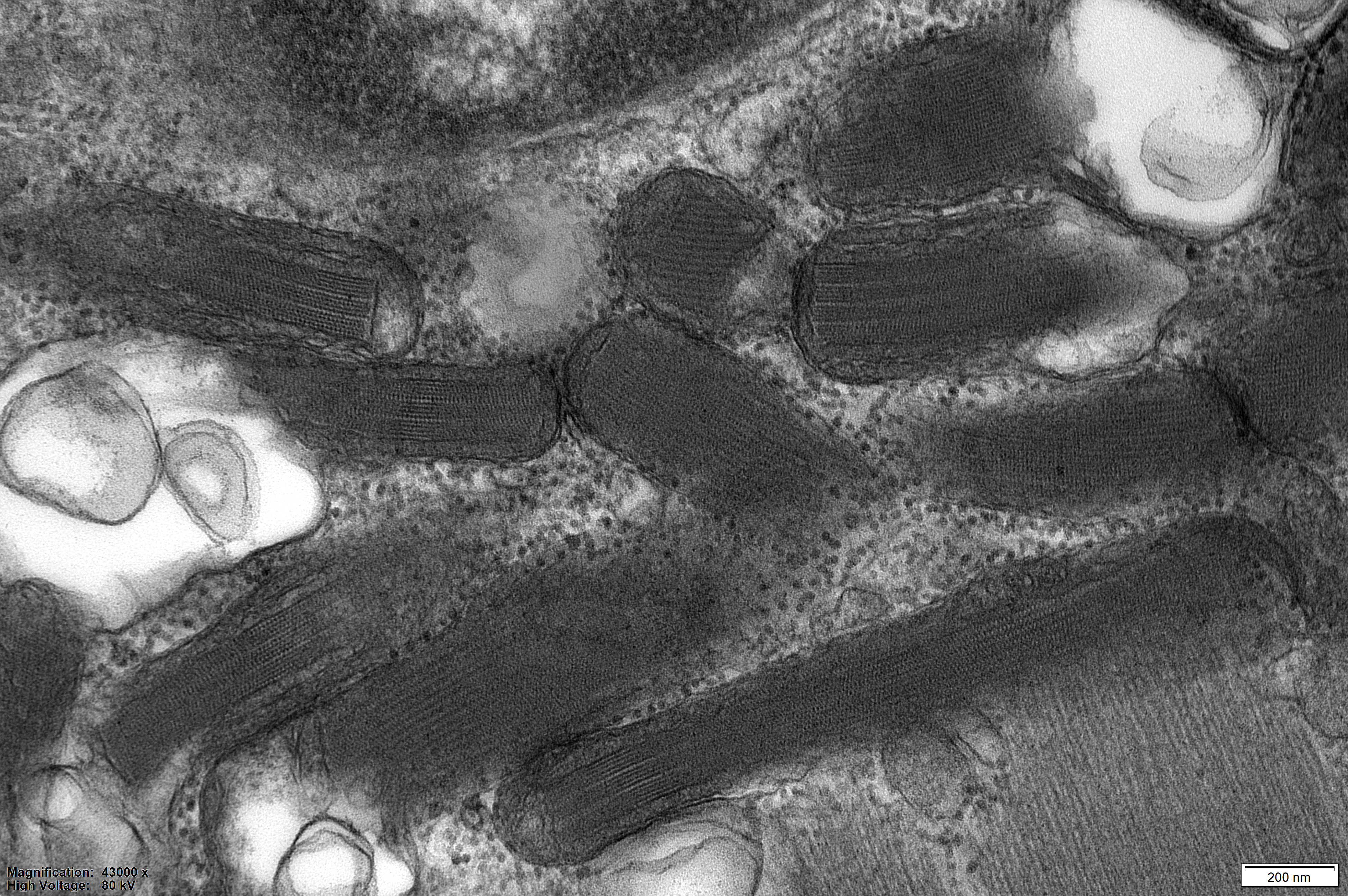
Ultrastructural Resolution of Different Mitochondrial Compartments
By Tasnim Arroum, Westfälische Wilhelms-University Münster
Inheritance
By Therese Kichuk, PhD Student in Molecular Biology at Princeton University
This image shows clusters of the budding yeast Saccharomyces cerevisiae. Mitochondria are shown in green and the endoplasmic reticulum is shown in red.
Reshaping of Mitochondria During Diabetes
By Licia Anna Pugliese and Luca Pesce, NEST ( National Enterprise for nanoScience and nanoTechnology),Scuola Normale Superiore
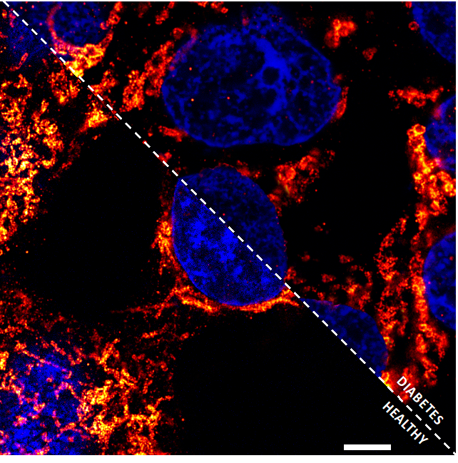
Confocal microscopy image showing cytokine-treated Ins-1E cells (on the right) and healthy cells (on the left) after staining with the specific marker for mitochondrial outer membrane TOM20 (red), and DAPI for nucleus (blue) to analyze the mitochondrial structure. The images were taken through a new super resolution method called Expansion Microscopy. Scale bar: 20 μm.
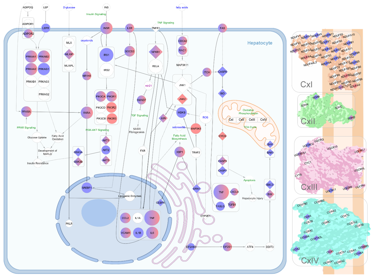
MiRNA-Dependent Regulation of NAFLD Pathogenesis with Emphasis on the Mitochondrial Aspect
By Maria Bograya, Center for Immunology and Cellular Biotechnology, Immanuel Kant Baltic Federal University
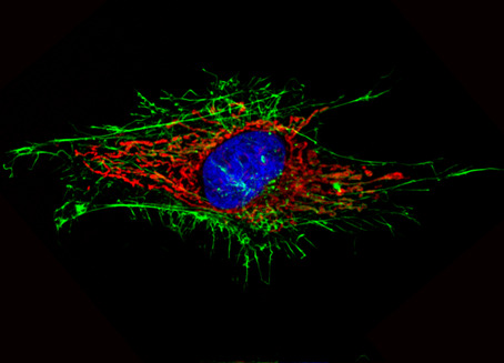
When Mitochondria Loves You Back
By Tanoy Dutta, Department of Chemistry, Indian Institute of Science Education and Research
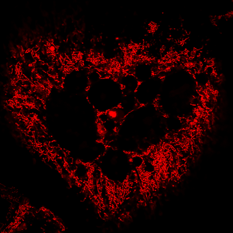
Confocal microscopy image showing a U-87 MG cell stained with a mitochondria-targeting small molecule organic fluorophore Quinaldine Red to study the mitochondrial dynamics
Mitochondrial Red Eye
by Agnieszka Fedoruk-Wyszomirska, Institute of Bioorganic Chemistry, Polish Academy of Sciences
Confocal microscopy image showing the mitochondrial network, cytoskeleton, and nucleus in MRC5 cell after FC treatment stained with MitoTracker Red CMXRos, Alexa Fluor 488 phalloidin, and Hoechst 33342, respectively.
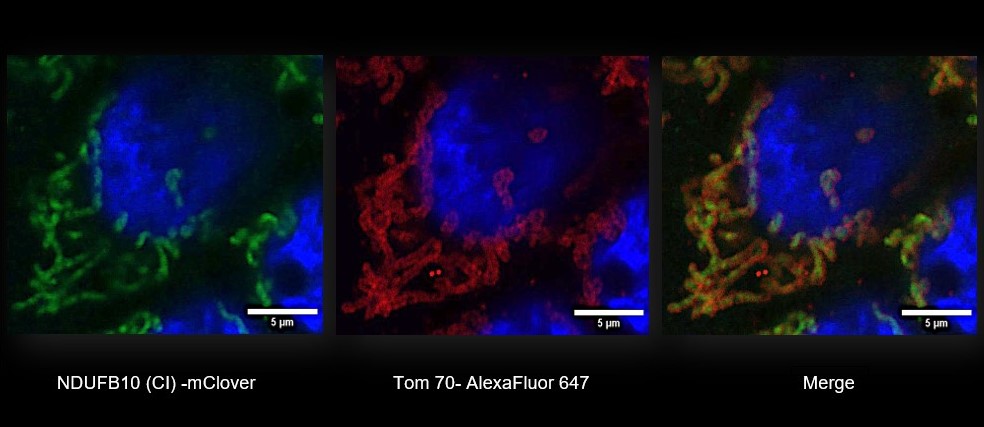
Uncovering Where the Mitochondrion Makes its Proteins
By Rolando Berlinguer Palmini and Matt Zorkau, Newcastle University
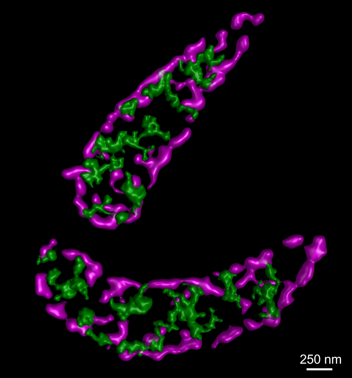
Synthesis of mitochondrial-encoded proteins in U20S cells was labelled with the methionine analogue HPG and detected by a fluorescent azide (green). Mitochondrial inner boundary membranes (magenta) were visualised by immunofluorescent labelling of Tim23. Stimulated emission depletion (STED) microscopy and surface rendering was used to determine sub-mitochondrial resolution of the targets.
MIRO1 Regulates Mitochondrial Trafficking in Cancer-Associated Fibroblasts
By Michael Cangkrama, ETH Zurich
MIRO1 labeling in the mitochondrial network of skin fibroblasts
Mitochondrion Before Oxaliplatin Storm
By Toni Martinez-Bernabe, University of Balearic Islands
Megacell
By Valentin Baumgartner, PhD student at the Laboratory for Urologic Oncology and Stem Cell Therapy, University Hospital Zurich
PC-3 prostate cancer cells were stained with TOM20 to analyse the mitochondrial network. Depicted here is one “Megacell” entangling other cells with its extensive mesh of mitochondria.
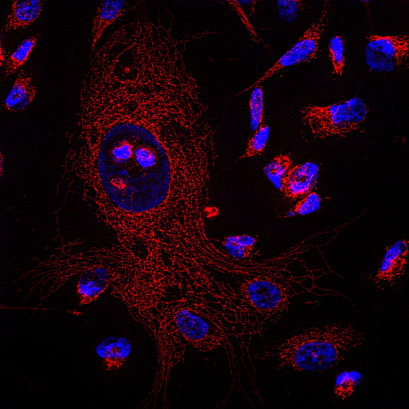
The Beauty & the Beast
By Arpit Mehrotra and Deepak Kumar Sharma, Council of Scientific & Industrial Research-Institute of Microbial Technology, India
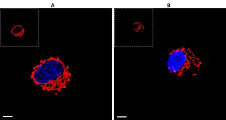
SH-SY5Y cells were stained with Mitotracker-RED dye (in Red) and Hoechst dye (in Blue) to track mitochondrial networking in control (A) and synuclein transfected (B) cells, wherein beauty of mitochondrial networking was observed in control and the synuclein mediated beastly-broken network was observed upon its overexpression. Scale bar-20µm
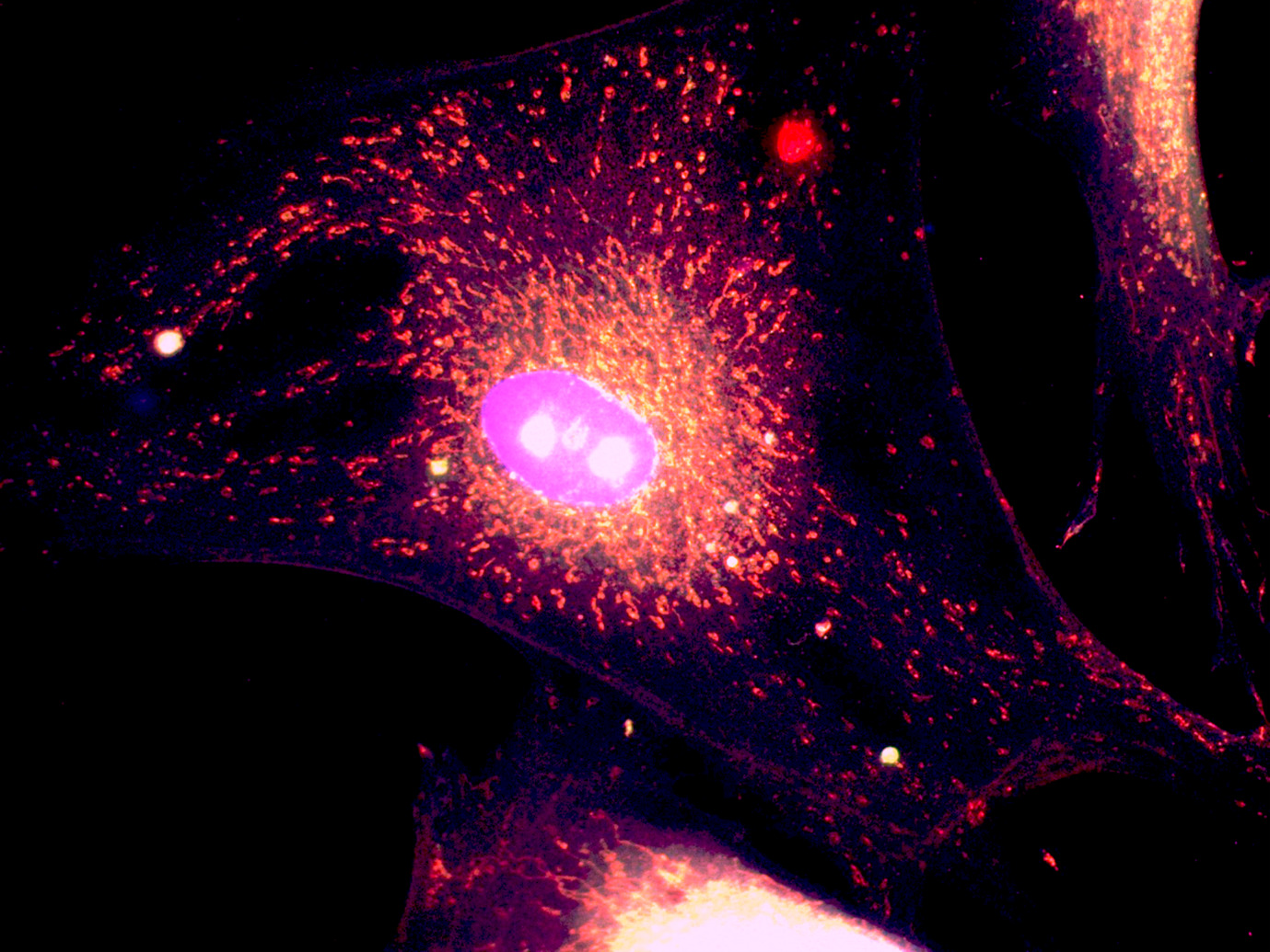
Mitochondria run parallel in mature cardiomyocytes
By Erminia Donnarumma, Institut Pasteur, France
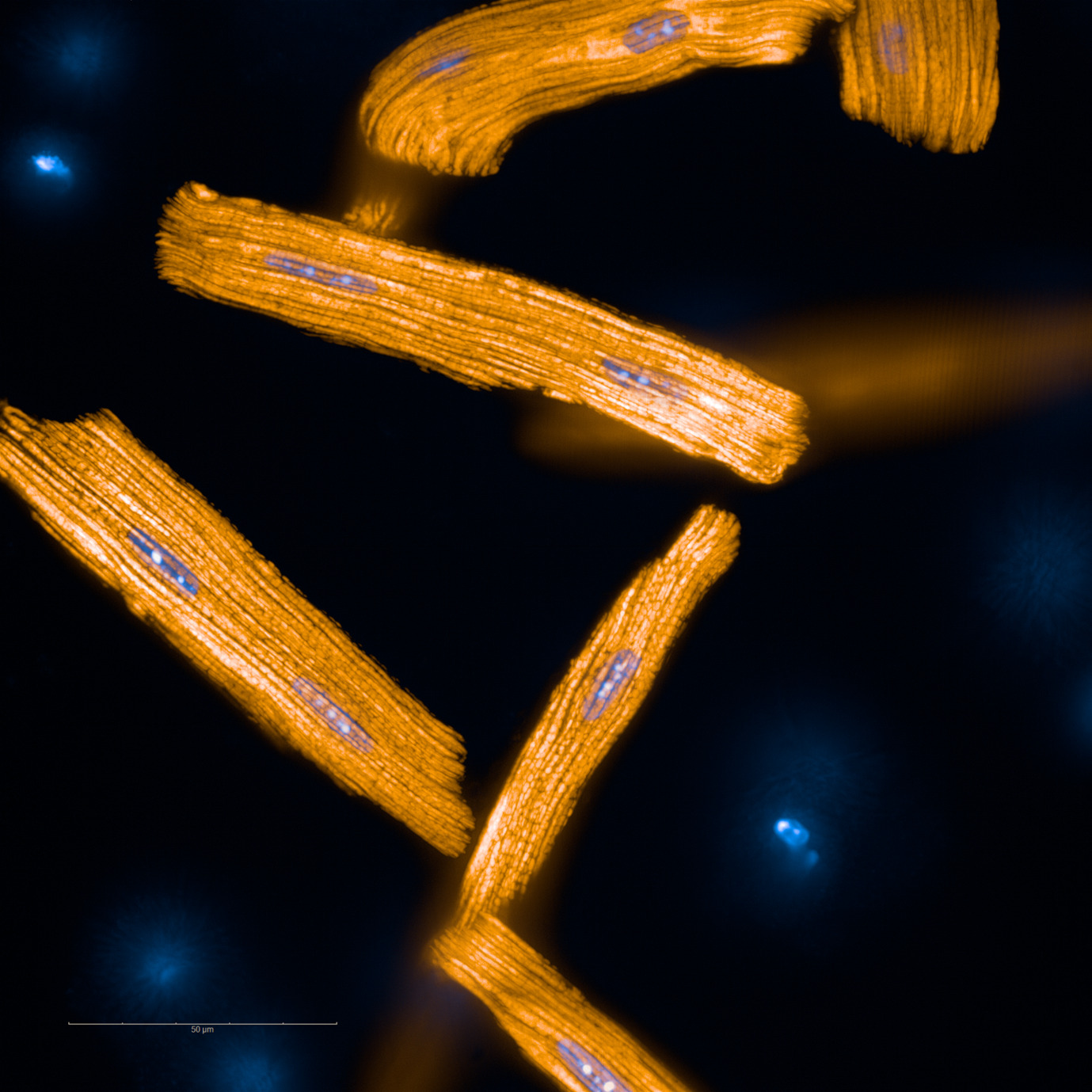
Adult primary cardiomyocytes (mouse) stained with TMRE (mitochondria, orange) and NucBlue (nuclei, blue).
Targeting Mitochondria 2022 Congress
October 26-28, 2022 - Berlin, Germany
wms-site.com