Microscopy Enables Detailed Insights into Mitochondria
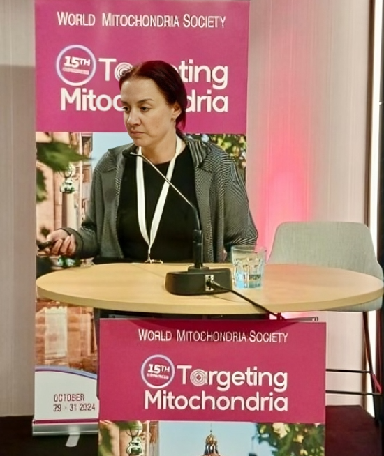
The scientific committee of Targeting Mitochondria World Congress invited Dr. Martin Kerschensteiner to present his excellent work.
A new microscopy technique combining confocal and two-photon excitation microscopy with in situ pharmacological and genetic manipulation has given researchers insight into how the nervous system responds to disease and injury at the mitochondrial level.
Reactive oxygen species are important intracellular signaling molecules, but their mode of action is complex: In low concentrations, they regulate key aspects of cellular function and behavior; at high concentrations, they can cause oxidative stress, which damages organelles, membranes and DNA.
The new method allows researchers to record the oxidation states of individual mitochondria with high spatial and temporal resolution, and to analyze how reduction/oxidation (redox) signaling unfolds in single cells and organelles in real time. 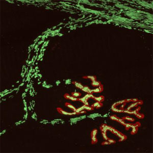
The nerve-cell mitochondria were imaged with a fluorescent redox sensor. Shown here is a peripheral nerve with the neuromuscular endplates stained in red. Courtesy of Ludwig Maximilian University and the Technical University of Munich.
"Redox signals have important physiological functions, but can also cause damage, for example, when present in high concentrations around immune cells," said professor Dr. Martin Kerschensteiner of Ludwig Maximilian University of Munich (LMU) and SyNergy, the Munich cluster for Systems Neurology. His team collaborated with that of Technical University of Munich (TUM) professor Dr. Thomas Misgeld, also of SyNergy.
In previous studies, Kerschensteiner and Misgeld found that oxidative damage of mitochondria could contribute to axonal destruction in inflammatory diseases such as multiple sclerosis.
For this work, they used redox-sensitive variants of green fluorescent protein (GFP) as visualization tools. They combined these with other biosensors and dyes, which allowed simultaneous monitoring of the redox signals and mitochondrial calcium currents, along with changes in electrical potential and pH gradient across the mitochondrial membrane.
The researchers used the method to study, for the first time, redox signal induction in response to neural damage in the mammalian nervous system (in this case, spinal cord injury). They found that severance of an axon results in a wave of mitochondrial oxidation beginning at the site of damage and propagating along the fiber. In addition, an influx of calcium at the axonal resection site was essential to cause functional damage to mitochondria.
"This appears to be a fail-safe system that is activated in response to stress and temporarily attenuates mitochondrial activity," Misgeld said. "Under pathological conditions, the contractions are more prolonged and may become irreversible, and this can ultimately result in irreparable damage to the nerve process."
Another first-ever result was the imaging of spontaneous contractions of mitochondria accompanied by a rapid shift in the redox state of the organelle.
Dr. Martin Kerschensteiner will present his strategic studies during Targeting Mitochondria 2014.
For more information: www.targeting-mitochondria.com


