
Meet the nominees for the WMS Best Scientific Image Award 2026!
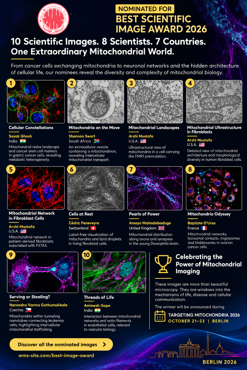
10 Scientific Images. 8 Scientists. 6 Countries.
One Extraordinary Mitochondrial World.
Meet the nominees for the WMS Best Scientific Image Award 2026!
Can a single scientific image change how we understand mitochondria?
From cancer cells exchanging mitochondria to neuronal networks, extracellular vesicles and the hidden architecture of cellular life, our nominees reveal the diversity and complexity of mitochondrial biology.
The World Mitochondria Society is pleased to present the 10 nominated images.
1. Sayak Ghosh | India
Cellular Constellations
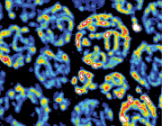
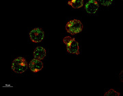
Exploring mitochondrial oxidative stress and the cancer stem cell marker CD44 in gastric cancer cells, revealing the metabolic heterogeneity of tumour populations.
2. Shannon Swart | South Africa
Mitochondria on the Move
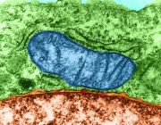
An electron microscopy image revealing a mitochondrion enclosed within an extracellular vesicle, opening questions about mitochondrial transport and intercellular communication.
3. Arshi Mustafa | USA
Mitochondrial Landscapes
An ultrastructural view of mitochondria in a cellular model carrying the FMR1 premutation, relevant to understanding mitochondrial biology in fragile X-associated disorders.
4. Arshi Mustafa | USA
Transmission Electron Microscopy of Mitochondrial Ultrastructure in Fibroblasts
Exploring the internal architecture and morphological diversity of mitochondria within human fibroblast cells.
5. Arshi Mustafa | USA
Mitochondrial Network in Fibroblast Cells
Visualizing mitochondrial networks in patient-derived fibroblasts associated with fragile X-associated tremor/ataxia syndrome (FXTAS).
6. Cédric Peneveyre | Switzerland
Cells at Rest
A label-free view of living fibroblasts, revealing mitochondria and lipid droplets without fluorescent staining.
7. Amaya Malmalabaduge | United Kingdom
Pearls of Power
Revealing the remarkable distribution of mitochondria along axons and synaptic terminals in the young Drosophila brain.
8. Baptiste D’Urso | France
Mitochondria Odyssey
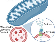
A fascinating composite image exploring mitochondrial networks, lysosomal contacts, migrasomes and blebbisomes in ovarian cancer cells.
9. Narendra Varma Gottumukkala | Czechia
Serving or Stealing?
Capturing mitochondrial structures within tunneling nanotubes connecting leukemia cells, highlighting an emerging mechanism of intercellular mitochondrial trafficking.
10. Animesh Gope | India
Threads of Life
Visualizing the relationship between mitochondrial networks and actin filaments in endothelial cells, central to mitochondrial dynamics and vascular biology.
These images are more than beautiful microscopy. They are windows into the mechanisms of life, disease and cellular communication.
Congratulations to all our nominees for their scientific creativity and contribution to mitochondrial imaging.
The WMS Best Scientific Image Award 2026 will be presented during the 17th Targeting Mitochondria Congress, October 21–23, in Berlin.
Explore all 10 images and their scientific stories: https://wms-site.com/best-image-award
For more information