Submit your work for the Best Image Award 2026


- Details
- Published on 20 July 2026

Best Scientific Image Award 2026: Show the World Your Mitochondrial Discovery
Can a single image change the way we see mitochondrial biology?
The World Mitochondria Society (WMS) is pleased to announce the opening of submissions for the Best Scientific Image Award 2026, an international competition celebrating the most outstanding scientific images in mitochondrial research.
From stunning microscopy to innovative scientific illustrations and AI-assisted visualizations, this award recognizes images that combine scientific excellence with visual impact. Every image tells a story, reveals a discovery, and helps communicate the remarkable complexity of mitochondria.
The winner will be announced during the Awards Ceremony of the 17th World Congress on Targeting Mitochondria, to be held in Berlin, Germany, October 21–23, 2026.
Who can participate?
The competition is open to researchers, clinicians, postdoctoral fellows, PhD students, engineers, and imaging specialists from academia and industry worldwide.
What can you submit?
We welcome original scientific images related to mitochondrial biology, including:
- Fluorescence and confocal microscopy
- Electron microscopy
- Live-cell imaging
- Super-resolution microscopy
- Clinical and translational imaging
- Scientific illustrations
- AI-assisted scientific visualizations
How to submit:
Each submission should include a high-resolution image, a title, a short scientific description (maximum 250 words), and the imaging technique used.
mitochondria[at]wms-site.com
Awards
The Best Scientific Image Award 2026 winner will receive:
- An official World Mitochondria Society Award Certificate
- Free registration to the 18th World Congress on Targeting Mitochondria 2027
- Recognition during the Awards Ceremony in Berlin
- International visibility through the WMS website, newsletter, and social media
- Inclusion in the WMS Scientific Image Gallery
New for 2026: People's Choice Award
For the first time, congress participants will vote for their favorite image during the meeting. This special award will recognize the image that best inspires and communicates mitochondrial science to the international community.
Submit Your Image
Submission deadline: October 12, 2026
Whether your work explores mitochondrial dynamics, metabolism, signaling, aging, disease, or therapeutic innovation, we encourage you to share the image that best represents your science.
One image can inspire a discovery. One image can inspire a community. We look forward to seeing yours.
Among the submission for BEST MITOCHONDRIA IMAGE 2026
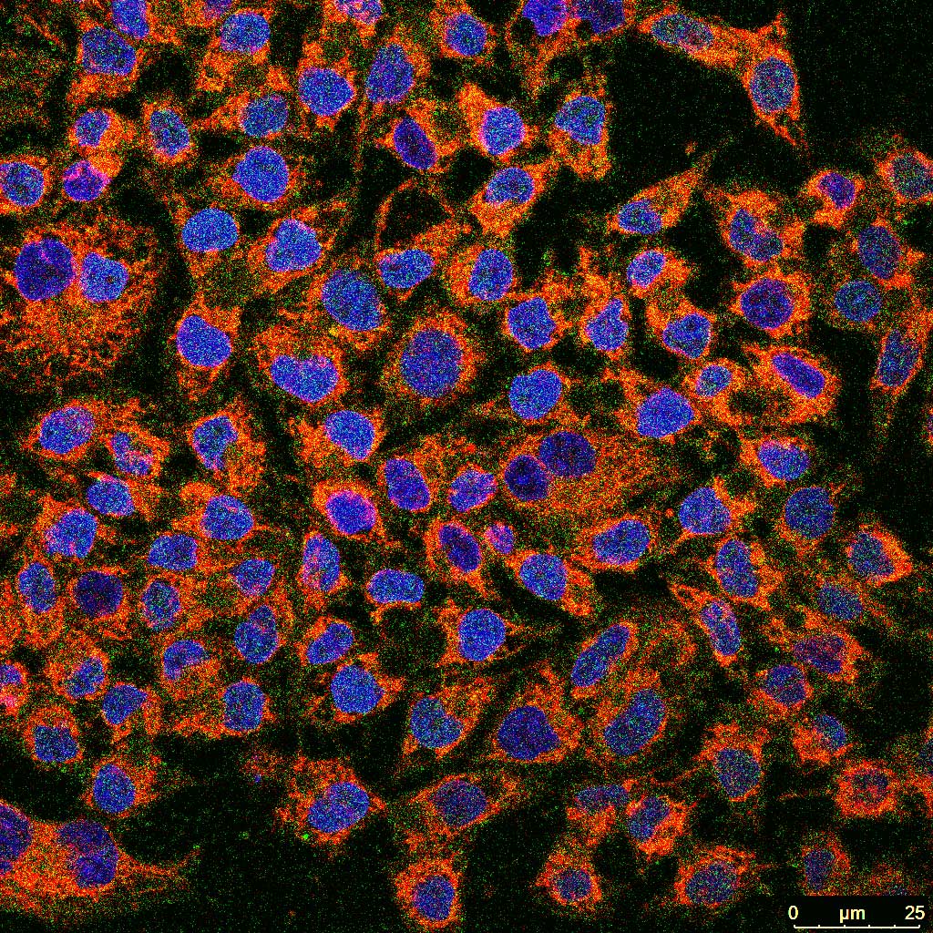
Image 1: Cellular Constellations: Illuminating the Mitochondrial Redox Landscape of Gastric Cancer
By Sayak Ghosh, M.Sc., Amity University Kolkata, India
Download Full Size image (TIF)
Description : Cancer cells undergo profound metabolic remodeling to sustain their rapid growth and survival under stressful conditions. This confocal fluorescence image illustrates the spatial relationship between mitochondrial reactive oxygen species (mtROS) production and the cancer stem cell marker CD44 in gastric adenocarcinoma cells. Cell nuclei are stained with DAPI (blue), MitoSOX™ Red (red) specifically labels mitochondrial superoxide, a key indicator of mitochondrial oxidative stress, and CD44 immunofluorescence (green) identifies cells with stemness-associated characteristics.
The heterogeneous distribution of mitochondrial superoxide and CD44 expression highlights the metabolic diversity that exists within the tumor cell population. Elevated mtROS has emerged as an important regulator of cancer cell plasticity, stemness, therapeutic resistance, and metastatic potential by modulating mitochondrial function and redox-sensitive signaling pathways. Simultaneously, CD44- positive cells represent a subpopulation with enhanced tumor-initiating capacity and resistance to conventional therapies. Visualizing these molecular events at the single-cell level provides valuable insight into the interplay between mitochondrial dysfunction and cancer stem cell biology.
This image underscores the importance of mitochondrial metabolism as a central determinant of tumor progression and demonstrates how advanced fluorescence microscopy can reveal the intricate cellular architecture underlying cancer biology. Such visualization contributes to understanding the molecular mechanisms that may guide the development of mitochondria-targeted therapeutic strategies for gastric cancer.
Imaging Technique Used
Immunocytochemistry (ICC) combined with MitoSOX™ Red staining and DAPI nuclear
counterstaining, imaged using a Leica TCS SP8 Laser Scanning Confocal Microscope.
Fluorescent markers:
Red: MitoSOX™ Red (mitochondrial superoxide)
Green: CD44 (cancer stem cell marker)
Blue: DAPI (cell nuclei).
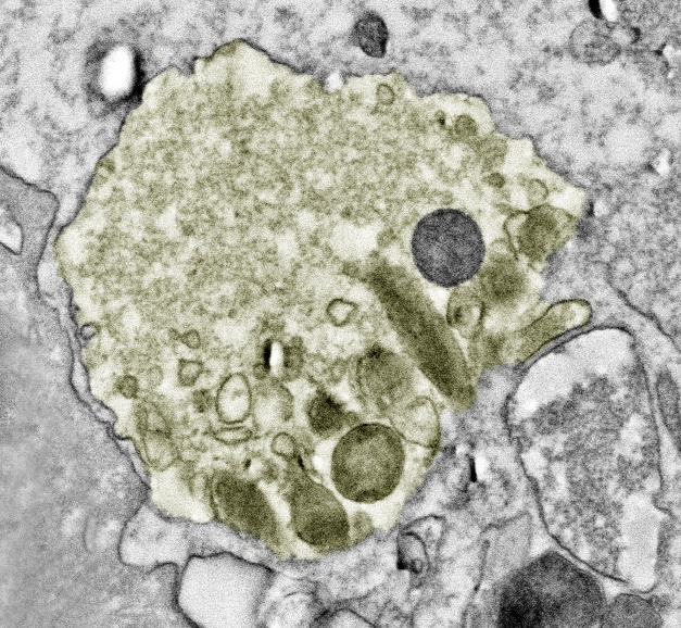
Image 2: Mitochondria on the move: Characterising extracellular mitochondria using electron microscopy
By Shannon Swart, PhD, Department of Physiological Sciences, University of Stellenbosch, South Africa
Download Full Size image (TIF)
Description: Mitochondria are traditionally regarded as intracellular organelles that serve as central hubs for energy production, redox homeostasis, and programmed cell death. However, under a range of physiological and pathophysiological conditions, mitochondria can translocate to the extracellular environment, either as free organelles or encapsulated within extracellular vesicles.
This electron microscopy image shows an extracellular vesicle containing a mitochondrion alongside other cellular components as cargo. The mitochondrion is distinctly identifiable by its characteristic double membrane and internal cristae. Extracellular mitochondria can originate from diverse cell types and may function as danger signals, mediators of intercellular communication, or functional organelles capable of supporting neighbouring cells with impaired mitochondrial function.
Consequently, extracellular mitochondria are emerging as important biomarkers of health and disease. Electron microscopy enables their precise identification and ultrastructural characterisation, offering valuable insights into mitochondrial biology.
Method: Scanning transmission electron microscopy using an Apreo Volumescope (ThermoFisher, Netherlands) and image acquisition was conducted with ThermoFisher Scientific's xt Microscopy software. Conducted at the Central Analytical Facility at Stellenbosch University.
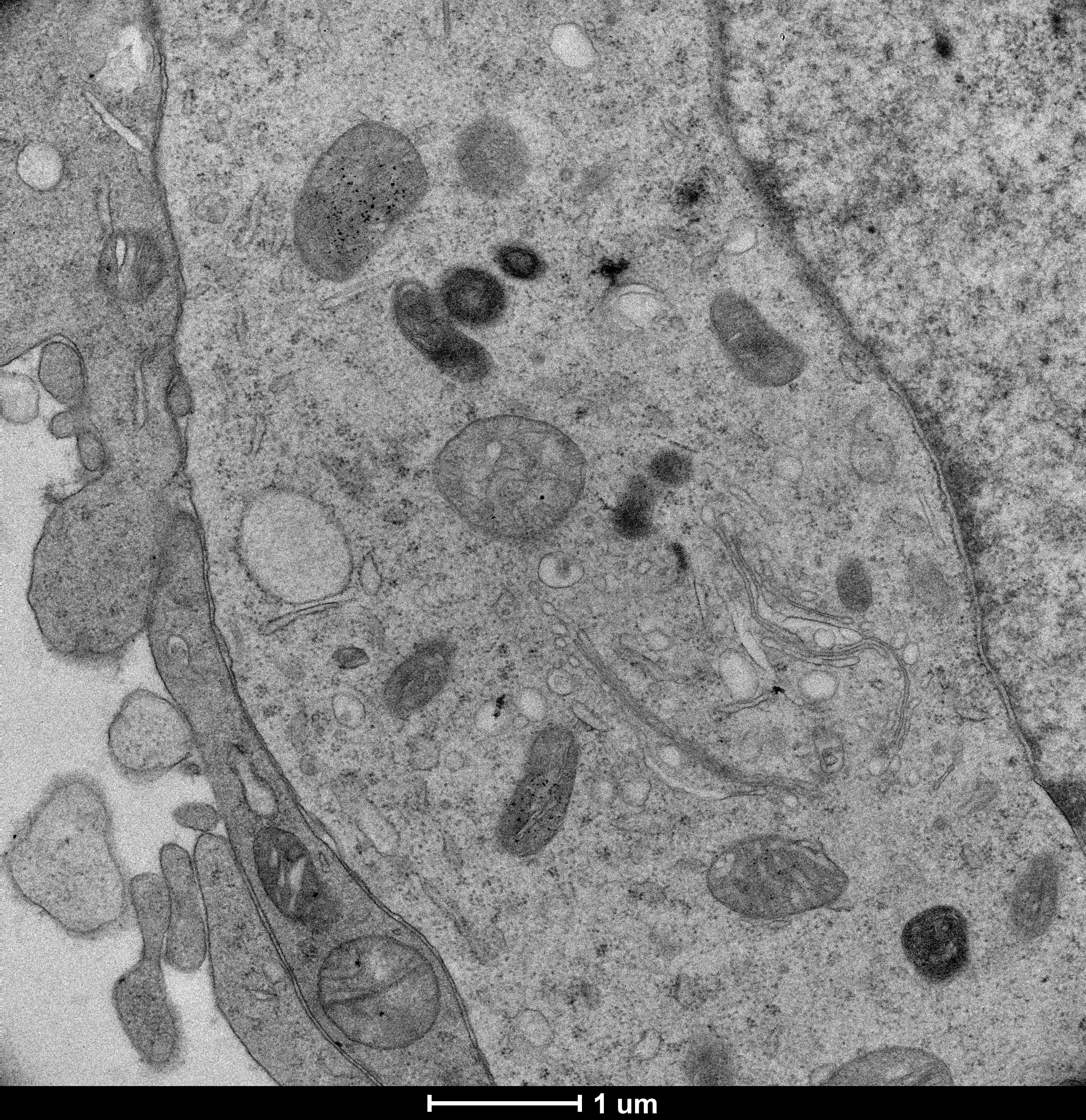
Image 3: Mitochondrial Landscapes: The Hidden Architecture of a Cell Carrying the FMR1 Premutation
By Arshi Mustafa, University of California, Davis, U.S.A.
Download Full Size image (TIF)
Description: This transmission electron microscopy image captures the mitochondrial landscape of an SKNMC cell expressing 95 CGG repeats in the FMR1 gene. Multiple mitochondria are distributed throughout the cytoplasm, revealing their characteristic morphology and intricate internal ultrastructural organization. The high-resolution image provides a detailed view of mitochondrial architecture within a human cell carrying the FMR1 premutation.
Study Context: SKNMC cells expressing 95 CGG repeats serve as a cellular model for investigating the effects of the FMR1 premutation on cellular and mitochondrial biology. This study uses ultrastructural imaging to examine mitochondrial morphology and organization, providing insight into how mitochondria are structured within cells carrying an expanded CGG repeat.
Method: Transmission electron microscopy (TEM) was used to visualize mitochondrial ultrastructure in SKNMC cells expressing 95 CGG repeats. Image acquired with a 1 µm scale bar. (University of California, School of Medicine, Dept. of Biochemistry and Molecular Medicine, U.S.A.).
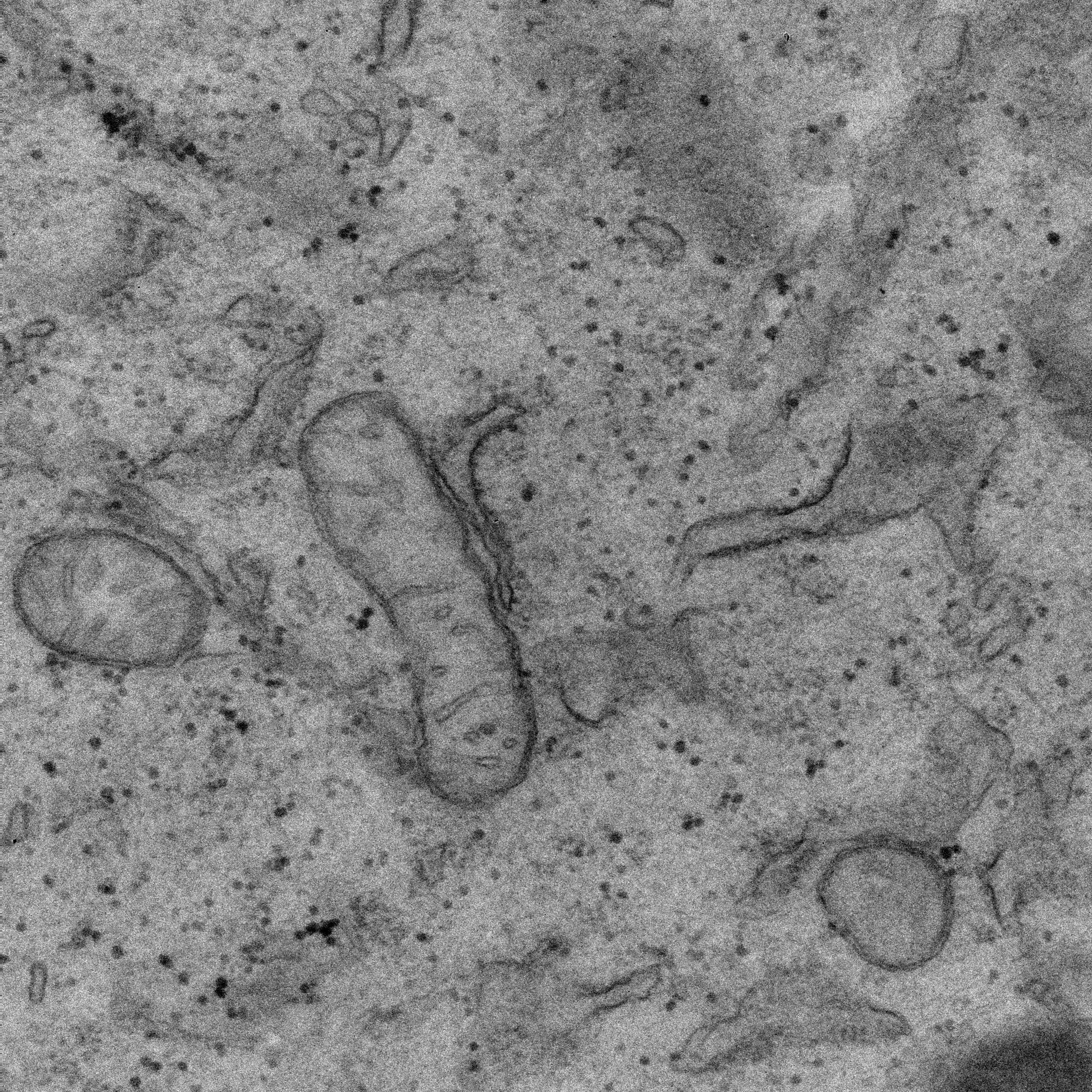
Image 4: Transmission Electron Microscopy of Mitochondrial Ultrastructure in Fibroblasts
By Arshi Mustafa, University of California, Davis, U.S.A.
Download Full Size image (TIF)
Description: This transmission electron microscopy image captures the mitochondrial landscape of a human fibroblast cell. Multiple mitochondria are distributed throughout the cytoplasm, revealing their characteristic morphology and internal ultrastructural organization. The high-resolution image provides a detailed view of mitochondrial architecture within a human cell.
Study Context: Fibroblast cells provide a valuable cellular model for investigating mitochondrial structure and function. This study uses ultrastructural imaging to examine mitochondrial morphology and organization, providing insight into the structural features of mitochondria at the nanoscale.
Method: Transmission electron microscopy (TEM) was used to visualize mitochondrial ultrastructure in fibroblast cells. The image reveals distinct mitochondrial profiles and their surrounding cytoplasmic environment. (University of California, School of Medicine, Department of Biochemistry and Molecular Medicine, U.S.A.)
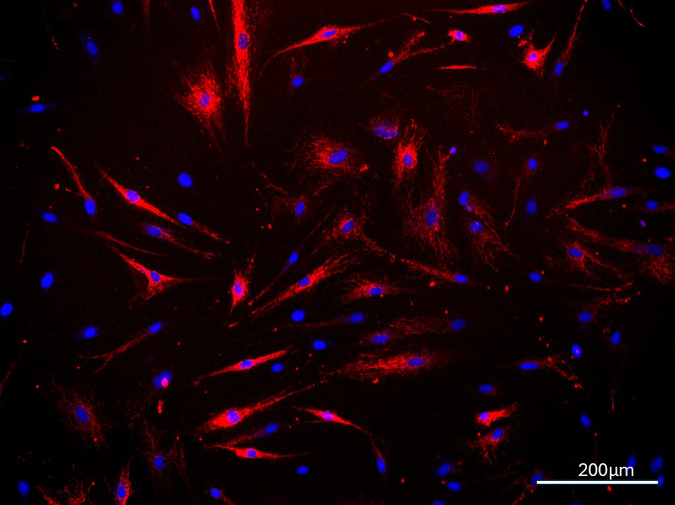
Image 5: Mitochondrial Network in Fibroblast Cells
By Arshi Mustafa, University of California, Davis, U.S.A.
Download Full Size image (TIF)
Description: This fluorescence microscopy image captures the mitochondrial landscape of patient-derived fibroblast cells from an individual with fragile X–associated tremor/ataxia syndrome (FXTAS). TOM20 immunostaining (red) reveals an extensive and intricate mitochondrial network distributed throughout the cytoplasm, while nuclei are visualized in blue. The image highlights the organization and complexity of mitochondria within human cells affected by FXTAS, neurodegenerative disorder.
Study Context: Patient-derived fibroblasts provide a human cellular model for investigating the effects of the FMR1 premutation associated with FXTAS on cellular and mitochondrial biology. Visualization of the mitochondrial network provides an opportunity to examine mitochondrial organization and morphology in patient-derived cells and to identify potential cellular features associated with disease.
Method: Immunofluorescence staining for TOM20 was used to visualize mitochondria in FXTAS patient-derived fibroblasts. Nuclei were counterstained with DAPI. Images were acquired using an EVOS fluorescence imaging system. Scale bar: 200 µm. (University of California, School of Medicine, Department of Biochemistry and Molecular Medicine, U.S.A.)
Image 6: Cells at rest
By Cédric Peneveyre, Nanolive SA, Switzerland
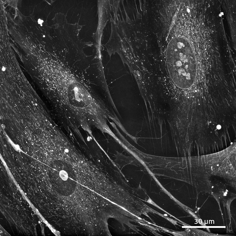
Description: Hundreds of mitochondria and lipid droplets fill these fibroblast cells, gathering densely around the cell nuclei, and stretching to their farthest tips. These optically dense organelles are made visible by their high refractive index properties. Simultaneous visualization of cytoplasm and cell membranes reveals areas of overlap and contact between cells, where cytoplasm stretches thin, and thick tubes trail like roots far from the cell body.
This image was taken 38 hours into a label-free timelapse, and the mitochondria in these cells appear to be undisturbed and at equilibrium. While fragmented or swollen mitochondria would indicate cell stress, the majority here are unbranched, of medium to long length, without swelling. Non-invasive imaging methods like interferometric holotomography are key to understanding the true nature of mitochondria, which are highly sensitive to perturbation by chemical dyes and phototoxicity.
Method: MRC-5 cells were plated and imaged for 38 hours using the Cell Explorer 96focus (Nanolive SA, Switzerland). FoV: 162.4 x 162.4 µm. This imaging technique is interferometric holotomography, which produces quantitative images without fluorescent labels or dyes.
Image 7: Pearls of Power: Mitochondrial Distribution Along Axons and Synapses of the Young Drosophila Brain
By Amaya Malmalabaduge, PhD Student, Natalia Sanchez-Soriano Lab, Department of Biochemistry and Systems Biology, University of Liverpool, UK
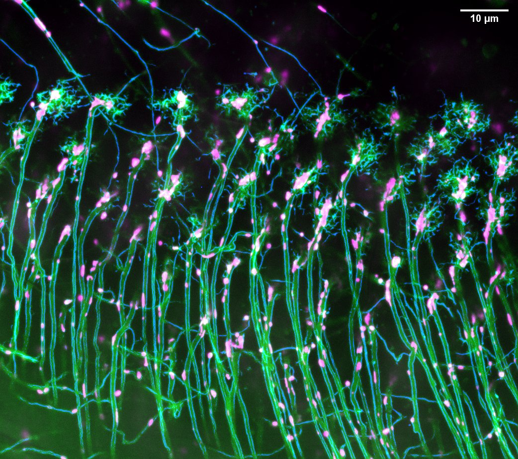
Description: Neurons are among the longest-lived and most energy-demanding cells and depend on the precise positioning of their mitochondria. This confocal image of T1 neurons in the optic lobe of a young (4–7 day old) Drosophila brain reveals mitochondria distributed along axons and within synaptic terminals. Neuronal membranes are visualised in green (UAS-myr::tdTomato) and mitochondria in magenta (UAS-mito-GFP), driven by GMR31F10-Gal4. Parallel axons carry mitochondria as discrete units, which accumulate as dense clusters within branched synaptic terminals. This distribution reflects function. Mitochondria supply ATP throughout the axon, powering ion pumps, motor proteins and cytoskeletal maintenance, with demand peaking at synapses, where they also fuel vesicle cycling and buffer calcium influx.
Mitochondrial positioning depends on the microtubule cytoskeleton. Kinesin and dynein motors, coupled via the Miro–Milton complex, transport mitochondria along axonal microtubules; fission and fusion remodel and segregate damaged components; and mitophagy delivers them to autophagosomes. However, microtubule dynamics and transport also depend on mitochondrial ATP, while excessive ROS from dysfunctional mitochondria may compromise microtubule integrity. During ageing, microtubules, autophagy and mitochondria decline in parallel, yet whether they fail independently or reinforce one another is unknown. This image shows the young, healthy brain: the baseline against which age-related fragmentation, stalled transport and impaired mitophagy are measured, and the state that therapies for neurodegeneration aim to restore. A single image highlights the central role of mitochondrial number, morphology and positioning, their interdependence with neuronal structures in healthy brains, and that the changes of ageing cannot be understood without seeing both together.
Imaging Information:
Microscope: 3i Marianas Spinning Disk (CCI University of Liverpool)
Green: UAS-myr::tdTomato (Neuronal membranes)
Magenta: UAS-mito-GFP (Mitochondria)
Driver: GMR31F10-Gal4 (T1 Neurons)
Blue: Axonal membrane outlines, generated in Fiji by edge-enhancement filter of the membrane channel
Image 8: Mitochondria Odyssey : Blebbisomes, migrasomes and other intercellular mitochondrial transport vesicles
By Baptiste D’Urso, PhD, CY Cergy Paris University, France
Download Full Size image (TIF)

Short Scientific Description: Alterations in mitochondrial dynamic and metabolism are key mechanisms in the progression of cancer. This image highlights, in ovarian adenocarcinoma cells, the morphological diversity of the mitochondrial network (coral red) as well as contact sites with lysosomes (cyan). On closer inspection, autophagolysosomes can be discerned. A cell is followed by a type of extracellular vesicle (EVs) associated with cell dynamics, containing fissioned mitochondria: a migrasome. Other vesicles for intercellular mitochondrial transfer are visible: blebbisomes. These are large, adherent EVs (5–20 µm) with properties similar to those of cells: they are contractile, possess a cytoskeleton, are rich in mitochondria, but lack a nucleus. There are four of them in this image – can you spot them?
Technical Aspect: This composite image combines various view from experiments, such as those involving extracellular matrix engineering and the induction of mitophagy. Three dyes are used: LysoTracker™ (cyan), MitoTracker™ (coral red) and Hoechst (blue). The cells are imaged in vivo using a Zeiss LSM 900 Airyscan 2 microscope fitted with a 63x NA 1.4 oil immersion objective. A custom-made Fiji LUT featuring the colours of the World Mitochondria Society was used for the mitochondria.
Image 9: Serving or Stealing? Intercellular Mitochondrial Trafficking in Leukemia
By Narendra Varma Gottumukkala, MSc, CEITEC-Masaryk University, Brno, Czechia.
Download Full Size image (TIF)
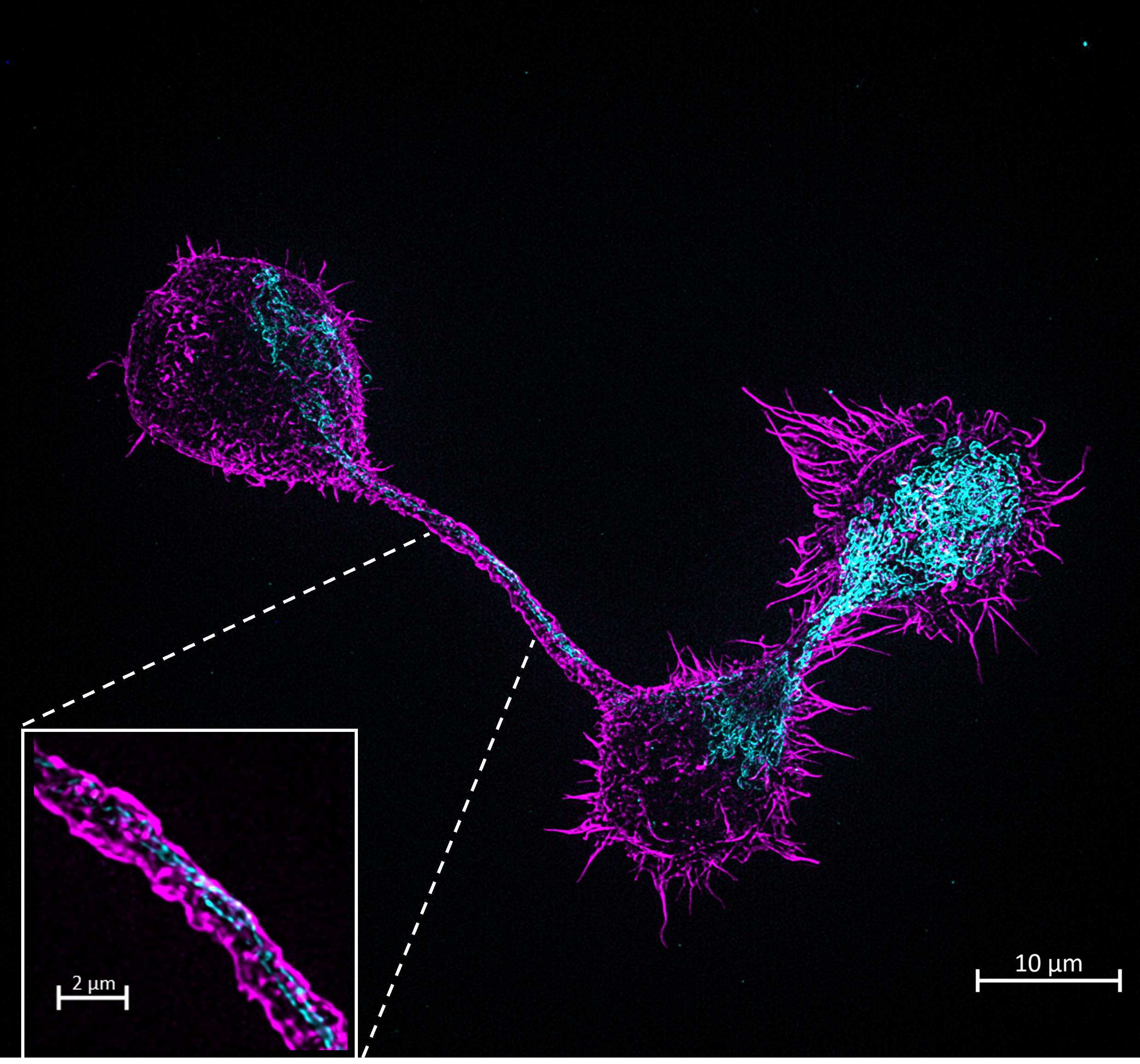
Scientific Description: In the hostile tumour microenvironment, mitochondria are the ultimate currency of survival. This super-resolution image captures two chronic lymphocytic leukemia (CLL) cells locked in a high-stakes metabolic exchange. Bridging them is a tunneling nanotube (TNT)- an ultra-thin F-actin conduit, stained in magenta (phalloidin) and threaded through its lumen is the cargo: mitochondria, labeled in cyan (TOMM20).
The image raises a fundamental question in cancer metabolism: are these cells serving or stealing? Intercellular mitochondrial transfer is an emerging hallmark of cancer adaptability. While cells can donate mitochondria to rescue damaged neighbours, leukemic cells weaponize the network- casting actin tethers to siphon healthy mitochondria from neighbouring cells, including immune effectors, fueling their own metabolic demands and driving disease progression while leaving donors energetically drained.
Technical Details: Visualizing organelles inside TNTs is notoriously difficult: at sub-micron diameters, conventional microscopy blurs the tube and its cargo into a single indistinguishable filament. Here, fixed CLL cells were immunostained for the mitochondrial outer-membrane protein TOMM20 (cyan) and F-actin (phalloidin, magenta) and imaged on a ZEISS Elyra 7 with Lattice SIM and a 63×/1.4 oil objective. The resolution gain of Lattice SIM resolves discrete mitochondrial puncta inside the nanotube lumen- cargo caught in transit. Complementary live-cell imaging confirms active mitochondrial trafficking through these conduits, establishing them as functional metabolic highways.
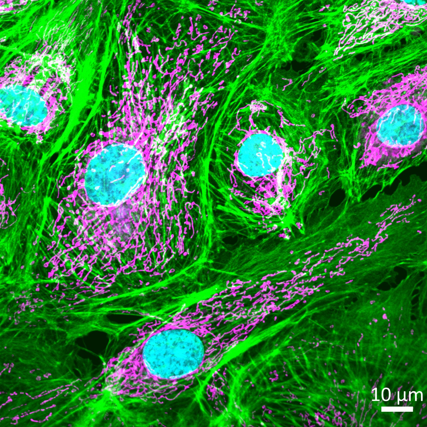
Image 10: Threads of life: Actin Filaments intersecting Mitoskeleton
By Animesh Gope, Division of Clinical Medicine, ICMR- Kolkata, India.
Download Full Size image (TIF)
Description of the image: The image highlights the mitochondrial network distributions. The outer mitochondrial membrane labelled with MitoTracker Red (Pseudo Violet) and Alexa Fluor 488 conjugated phalloidin (green), which target the intracellular mitochondrial network and cytoskeletal actin filaments. This interaction is crucial for mitochondrial dynamics (movement and division), and cellular responses to stress. In addition, the nucleus was stained with DAPI (Pseudo Turquoise) Scale Bar 10 μm.
Context of the study: It's an important model system to study the vascular function, endothelial barrier regulation and endothelial diseases like atherosclerosis and hypertension. Mitochondrial dysfunction in the arterial cell wall leads to formation of reactive oxygen species (ROS) which causes oxidative stress and damages the endothelial cells and hampers the normal vascular function. This ROS activates the inflammatory complex such as NLRP3 inflammasome and pro-inflammatory cytokines, such as IL-1β and IL-18 and accelerates plaque growth and promotes the apoptosis and necrosis of endothelial cells.
Method: Bovine Pulmonary Artery Endothelial cells were isolated and cultured in sterile environment, adding Dulbecco's modified eagle medium (DMEM) or minimum essential medium (MEM) as a supplements with 10% Fetal Bovine Serum (FBS) and antibiotic cocktail is added to prevent the bacterial contamination. The culture is maintained at 37°C and 5% CO₂.
Technical details: The images were captured by the inverted Axio observer confocal laser scanning microscope (Carl Zeiss LSM710) fitted with 63x (NA 1.4 Oil DIC III) objective lens and analysis by the Zen Blue software.
BEST MITOCHONDRIA IMAGE AWARD 2025 - FINALIST LIST
Cédric Peneveyre